RSNA Press Release
- Imaging of carotid artery plaque with MRI can accurately predict future cardiovascular events in people without a history of cardiovascular disease.
- Imaging results were compared with cardiovascular events, including heart attacks, stroke and death, for an average of 5.5 years after examination.
- Cardiovascular events occurred in 59 of the 946 patients in the study group.
Carotid Artery MRI Helps Predict Likelihood of Strokes and Heart Attacks
Released: March 4, 2014
| Media Contacts: | |
| RSNA Media Relations: | 1-630-590-7762 |
| Linda Brooks 1-630-590-7738 lbrooks@rsna.org |
Maureen Morley 1-630-590-7754 mmorley@rsna.org |
OAK BROOK, Ill. – Noninvasive imaging of carotid artery plaque with MRI can accurately predict future cardiovascular events like strokes and heart attacks in people without a history of cardiovascular disease, according to a new study published online in the journal Radiology.
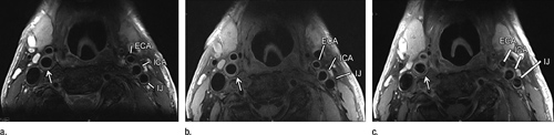
Researchers have long known that some arterial plaque is more dangerous because of its vulnerability to rupture. MRI can discern features of vulnerable plaque, such as a lipid core with a thin fibrous cap. This ability makes MRI a potentially valuable tool for identifying patients at risk for subsequent cardiovascular events.

To study the predictive value of MRI plaque imaging, researchers performed carotid artery ultrasound and MRI on 946 asymptomatic patients from the Multi-Ethnic Study of Atherosclerosis (MESA). The carotid arteries are the large vessels located on each side of the neck that carry oxygenated blood to the front part of the brain. They are highly accessible for imaging, and their condition tends to mirror that of the coronary arteries that supply the heart with oxygenated blood.
The researchers used ultrasound to assess carotid wall thickness and MRI to define carotid plaque composition and the remodeling index, a measure of changes in vessel size. Imaging results were compared with cardiovascular events, including heart attacks, stroke and death, for an average of 5.5 years after examination.
"We studied asymptomatic individuals with a low risk of cardiovascular events at baseline and used noninvasive imaging to predict the risk of an event downstream," said David A. Bluemke, M.D, Ph.D., from the National Institutes of Health Clinical Center in Bethesda, Md. "This is the first population-based prospective study to determine if vulnerable plaque features by MRI add to the risk of a cardiovascular event beyond the traditional risk factors."
Cardiovascular events occurred in 59 of the patients. Abnormal thickening of the carotid artery wall and the presence of a lipid core and calcium in the internal carotid artery on MRI were significant predictors of subsequent events. A lipid core was present in almost half of the patients who had an event, compared with only 17.8 percent of those who did not have an event.
"The primary factors that predicted future risk were measures of vessel wall thickness in combination with the presence or absence of a lipid core," Dr. Bluemke said. "The presence of a lipid core was 50 percent more common in people who had subsequent events."
Use of MRI improved the reclassification of baseline cardiovascular risk in the study group. When both the carotid remodeling index and lipid core were used for risk stratification, approximately 16 percent more patients with events and 7 percent without events were correctly reclassified compared with the use of traditional risk factors.
"The results bolster the use of MRI as a surrogate marker of efficacy in therapeutic studies and point to a role in determining which patients might need more aggressive treatments," Dr. Bluemke said. "As risk factor prediction gets better, we'll be able to screen more intelligently and use more intensive treatments in those individuals who face a higher risk of cardiovascular events."
Dr. Bluemke noted that sequences for plaque composition could be readily added to existing carotid MRI angiography protocols for clinical purposes.
"Carotid MRI has significant advantages," he said. "The results are more reproducible than those of ultrasound."
Availability and cost effectiveness could limit the use of MRI, Dr. Bluemke cautioned. However, ultrasound and computed tomography (CT) are viable alternatives for screening, he said, especially with improvements in CT dose reduction.
# # #
"Carotid Artery Plaque Morphology and Composition in Relation to Incident Cardiovascular Events: The Multi-Ethnic Study of Atherosclerosis (MESA)." Collaborating with Dr. Bluemke were Anna E. H. Zavodni, M.D., Bruce A. Wasserman, M.D., Robyn L. McClelland, Ph.D., Antoinette S. Gomes, M.D., Aaron R. Folsom, M.D., M.P.H., Joseph F. Polak, M.D., M.P.H., and João A. C. Lima, M.D.
Radiology is edited by Herbert Y. Kressel, M.D., Harvard Medical School, Boston, Mass., and owned and published by the Radiological Society of North America, Inc. (http://radiology.rsna.org/)
RSNA is an association of more than 53,000 radiologists, radiation oncologists, medical physicists and related scientists, promoting excellence in patient care and health care delivery through education, research and technologic innovation. The Society is based in Oak Brook, Ill. (RSNA.org)
For patient-friendly information on MRI, visit RadiologyInfo.org.
Images
|
High-res (TIF) version (Right-click and Save As) |
High-res (TIF) version (Right-click and Save As) |
High-res (TIF) version (Right-click and Save As) |


 PDF
PDF