RSNA Press Release
- A minimally invasive treatment for herniated — or "slipped" — disc gives patients significant long-term relief from sciatica pain.
- Conversely, patients treated only with pain relievers, anti-inflammatory medications and muscle relaxants, after initial remission, reported increased pain and decreased mobility a year later.
- In percutaneous disc decompression, a needle is used to puncture the disc, and tissue is removed or dissolved.
Outpatient Disc Treatment Gives Long-Term Back Pain Relief
Released: December 2, 2009
| Media Contacts: | RSNA Newsroom | 1-312-949-3233 |
| Before 11/28/09 or after 12/03/09: | RSNA Media Relations: | 1-630- 590-7762 |
| |
Linda Brooks 1-630-590-7738 lbrooks@rsna.org |
Maureen Morley 1-630-590-7754 mmorley@rsna.org |
CHICAGO — A randomized, controlled study comparing standard conservative therapy to a minimally invasive treatment called percutaneous disc decompression for painful herniated disc revealed that while both treatments help patients in the short run, only disc decompression kept patients pain free up to two years later. Results of the study, the first of its kind, were presented today at the annual meeting of the Radiological Society of North America (RSNA).
Discs are sponge-like cushions that rest between the bones of the spine. When a disc bulges or herniates outward, it can cause irritation or pressure on the spinal nerves, resulting in a condition known as sciatica. Sciatica is characterized by back and leg pain and weakness. Physicians often recommend that patients try six weeks of anti-inflammatory and pain medications before considering other treatments.
"Most protocols call for a minimal approach to initially treat a herniated disc," said Alexios Kelekis, M.D., Ph.D., assistant professor of interventional radiology at the University of Athens in Greece. "But by deflating the disc and giving the nerve root the space it needs, disc decompression solves the problem of root irritation and prevails in the long run."
In percutaneous disc decompression, interventional radiologists use image guidance to puncture a bulging disc through the skin with a needle and deflate the disc by either removing some tissue or using energy to dissolve it. The procedure is performed on an outpatient basis under local anesthesia, and patients are usually able to return to normal activities within 30 days.
In this study, Dr. Kelekis and colleagues treated two groups of patients with herniated discs and sciatica confirmed by MRI. Both groups included 17 men and 14 women complaining of back and leg pain. The mean age of patients was 36.
Both groups had tried different conservative treatments in the past without success. The first group, which received six weeks of rigorous conservative therapy consisting of analgesics, anti-inflammatory drugs and muscle relaxants, reported pain reduction and improved mobility at the end of treatment. The second group underwent percutaneous disc decompression.
Upon completion of treatment, patients in both groups were clinically evaluated and completed a questionnaire designed to assess pain relief, quality of life and mobility improvement at intervals of three months, 12 months and 24 months later.
Both patient groups reported pain reduction and increased mobility at the three-month interval. However, one year and two years after treatment, patients who had undergone disc decompression continued to improve, while patients who received only conservative therapy reported that their pain had returned and their mobility had decreased.
"Up until 12 months following therapy, both groups of patients were doing great," Dr. Kelekis said. "But by 12 months beyond treatment, patients who received only conservative therapy had returned to their initial pain levels."
Percutaneous disc decompression is suitable only for herniated discs that are not ruptured or too compressed.
Co-authors are Dimitrios Filippiadis, M.D., Dimitrios Erginousakis, Elias Brountzos, M.D., Nikolaos Ptohis, and Nikolaos Kelekis, M.D.
# # #
Note: Copies of RSNA 2009 news releases and electronic images will be available online at RSNA.org/press09 beginning Monday, Nov. 30.
RSNA is an association of more than 44,000 radiologists, radiation oncologists, medical physicists and related scientists committed to excellence in patient care through education and research. The Society is based in Oak Brook, Ill. (RSNA.org)
Editor's note: The data in these releases may differ from those in the printed abstract and those actually presented at the meeting, as researchers continue to update their data right up until the meeting. To ensure you are using the most up-to-date information, please call the RSNA Newsroom at 1-312-949-3233.
For patient-friendly information on image-guided therapies, visit RadiologyInfo.org.
| Abstract: |
Video clips
- WMV format
- Video clip (1,677 Kbyte)
A video showing cervical disc treatment. - Video clip (808 Kbyte)
A video showing treatment of a herniated disc in the lumbar region.
Images (.JPG format)
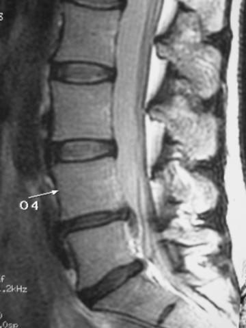 Figure 1. A magnetic resonance (MR) image of the spine prior to treatment for a herniated disc. High-res (TIF) version (Right-click and Save As) |
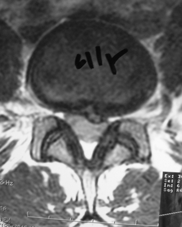 Figure 2. A magnetic resonance (MR) image of the spine prior to treatment for a herniated disc. High-res (TIF) version (Right-click and Save As) |
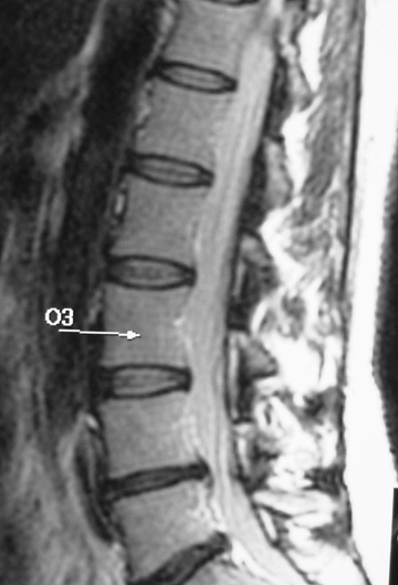 Figure 3. A magnetic resonance (MR) image of the spine one year after treatment for a herniated disc. High-res (TIF) version (Right-click and Save As) |
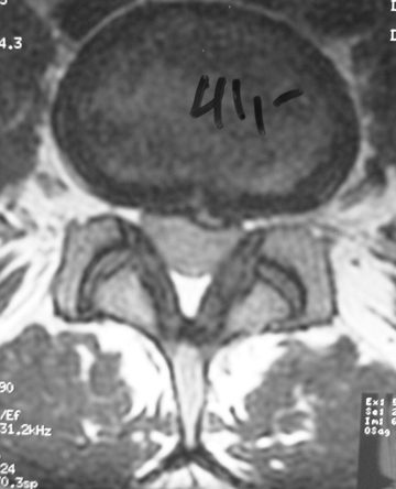 Figure 4. A magnetic resonance (MR) image of the spine one year after treatment for a herniated disc. High-res (TIF) version (Right-click and Save As) |
 PDF
PDF