RSNA Press Release
- Researchers are using a new robotic grip device to rehabilitate chronic stroke patients.
- This is the first study using fMRI to map the brain in order to track stroke rehabilitation.
- The study found that the brain is malleable for a longer period of time after stroke than previously thought, making permanent rehabilitation possible even after six months.
Robotic Technology Improves Stroke Rehabilitation
Released: December 3, 2008
| Media Contacts: | RSNA Newsroom | 1-312-949-3233 |
| Before 11/29/08 or after 12/04/08: | RSNA Media Relations: | (630) 590-7762 |
| |
Maureen Morley 1-630-590-7754 mmorley@rsna.org |
Linda Brooks 1-630-590-7738 lbrooks@rsna.org |
CHICAGO — Research scientists using a novel, hand-operated robotic device and functional MRI (fMRI) have found that chronic stroke patients can be rehabilitated, according to a study presented today at the annual meeting of the Radiological Society of North America (RSNA). This is the first study using fMRI to map the brain in order to track stroke rehabilitation.
"We have shown that the brain has the ability to regain function through rehabilitative exercises following a stroke," said A. Aria Tzika, Ph.D., director of the NMR Surgical Laboratory at Massachusetts General Hospital (MGH) and Shriners Burn Institute and assistant professor in the Department of Surgery at Harvard Medical School in Boston. "We have learned that the brain is malleable, even six months or more after a stroke, which is a longer period of time than previously thought."
According to the Centers for Disease Control and Prevention, stroke is the third leading cause of death in the U.S. and a principal cause of severe long-term disability. Approximately 700,000 strokes occur annually in the U.S., and 80 percent to 90 percent of stroke survivors have motor weakness.
Previously, it was believed that there was only a short window of three to six months following a stroke when rehabilitation could make an improvement.
"Our research is important because 65 percent of people who have a stroke affecting hand use are still unable to incorporate the affected hand into their daily activities after six months," Dr. Tzika said.
Dr. Tzika is an affiliated member of the Athinoula A. Martinos Center for Biomedical Imaging in the Department of Radiology at MGH, where the research is ongoing.
To determine if stroke rehabilitation after six months was possible, the researchers studied five right-hand dominant patients who had strokes at least six months prior that affected the left side of the brain and, consequently, use of the right hand.
For the study, the patients squeezed a special MR-compatible robotic device for an hour a day, three days per week for four weeks. fMRI exams were performed before, during, upon completion of training and after a non-training period to assess permanence of rehabilitation. fMRI measures the tiny changes in blood oxygenation level that occur when a part of the brain is active.
The results showed that rehabilitation using hand training significantly increased activation in the cortex, which is the area in the brain that corresponds with hand use. Furthermore, the increased cortical activation persisted in the stroke patients who had exercised during the training period but then stopped for several months.
"These findings should give hope to people who have had strokes, their families and the rehabilitative specialists who treat them," Dr. Tzika said.
Co-authors are Dionyssios Mintzopoulos, Ph.D., Azadeh Khanicheh, Ph.D., Bruce Rosen, M.D., Ph.D., Loukas Astrakas, Ph.D., and Michael Moskowitz, M.D.
# # #
RSNA is an association of more than 42,000 radiologists, radiation oncologists, medical physicists and related scientists committed to excellence in patient care through education and research. The Society is based in Oak Brook, Ill. (RSNA.org)
Editor's note: The data in these releases may differ from those in the printed abstract and those actually presented at the meeting, as researchers continue to update their data right up until the meeting. To ensure you are using the most up-to-date information, please call the RSNA Newsroom at 1-312-949-3233.
For patient-friendly information on fMRI, visit RadiologyInfo.org.
| Abstract: |
Images (.JPG format)
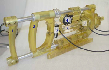 Figure 1. The second-generation prototype Magnetic Resonance Compatible Hand-Induced Robotic Device, or, MR_CHIROD.
(Right-click and Save As) |
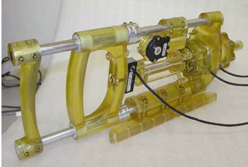 Figure 2. The second-generation prototype Magnetic Resonance Compatible Hand-Induced Robotic Device, or, MR_CHIROD. High-res (TIF) version (Right-click and Save As) |
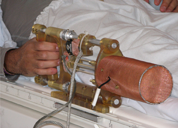 Figure 3. A volunteer squeezing the handles of the Magnetic Resonance Compatible Hand-Induced Robotic Device (MR_CHIROD) while lying in the magnet of the machine. High-res (TIF) version (Right-click and Save As) |
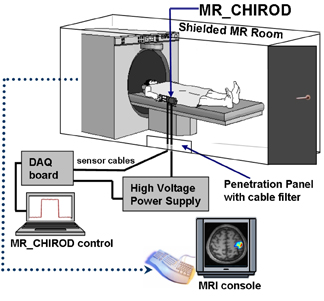 Figure 4. Diagram illustrating the concept of on-line brain mapping using fMRI and a Magnetic Resonance Compatible Hand-Induced Robotic Device (MR_CHIROD). High-res (TIF) version (Right-click and Save As) |
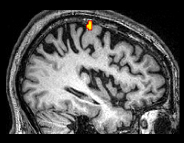 Figure 5. An fMRI image illustrating the area of the brain that corresponds with a patient's hand use before training. Effort level is 45percent of the patient's maximum hand-strength. High-res (TIF) version (Right-click and Save As) |
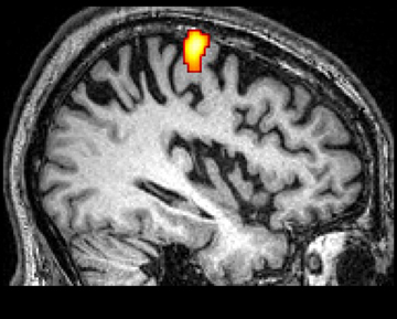 Figure 6. An fMRI image illustrating the area of the brain that corresponds with a patient's hand use before training. Effort level is 60percent of the patient's maximum hand-strength. High-res (TIF) version (Right-click and Save As) |
 Figure 7. An fMRI image illustrating the area in the brain that corresponds with a patient's hand use before training. Effort levels are 45percent (top) and 60percent (bottom) of the patient's maximum hand-strength. High-res (TIF) version (Right-click and Save As) |
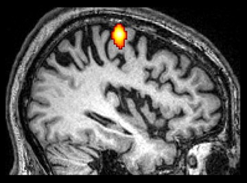 Figure 8. An fMRI image illustrating the area in the brain that corresponds with a patient's hand use after eight weeks of training. Effort level is 45percent of the patient's maximum hand-strength. High-res (TIF) version (Right-click and Save As) |
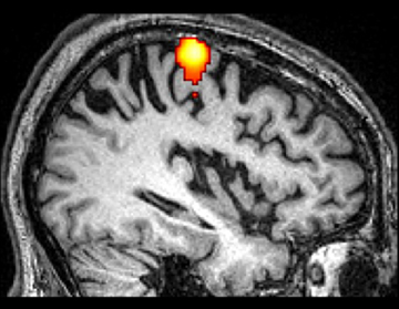 Figure 9. An fMRI image illustrating the area in the brain that corresponds with a patient's hand use after 8 weeks of training. Effort level is 60percent of the patient's maximum hand-strength. High-res (TIF) version (Right-click and Save As) |
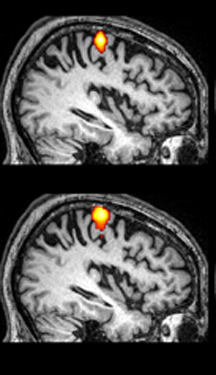 Figure 10. An fMRI image illustrating the area in the brain that corresponds with a patient's hand use after eight weeks of training. Effort levels are 45percent (top) and 60percent (bottom) of the patient's maximum hand-strength. High-res (TIF) version (Right-click and Save As) |
 Figure 11. An fMRI image illustrating the area in the brain that corresponds with a patient's hand use one month after training was completed. Effort level is 45percent of the patient's maximum hand-strength. High-res (TIF) version (Right-click and Save As) |
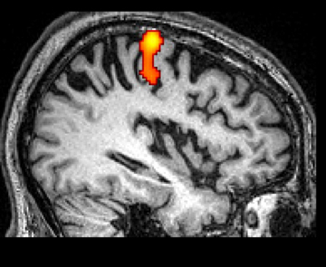 Figure 12. An fMRI image illustrating the area in the brain that corresponds with a patient's hand use one month after training was completed. Effort level is 60percent of the patient's maximum hand-strength. High-res (TIF) version (Right-click and Save As) |
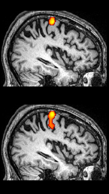 Figure 13. An fMRI image illustrating the area in the brain that corresponds with a patient's hand use one month after training was completed. Effort levels are 45percent (top) and 60percent (bottom) of the patient's maximum hand-strength. High-res (TIF) version (Right-click and Save As) |
 Figure 14. This fMRI image shows the area of the brain that corresponds with a patient's hand use before training, after eight weeks of training, and one month after the patient completed training. Effort levels are at 45percent (top) and 60percent (bottom). High-res (TIF) version (Right-click and Save As) |
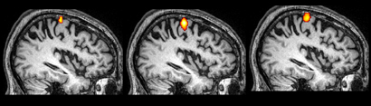 Figure 15. This fMRI image illustrates the area in the brain that corresponds with hand use of a patient before training (left), after eight weeks of training (middle), and one month after training was completed (right) at a 45percent effort level. High-res (TIF) version (Right-click and Save As) |
 Figure 16. This fMRI image illustrates the area in the brain that corresponds with hand use of a patient before training (left), after eight weeks of training (middle), and one month after training was completed (right) at a 60percent effort level. High-res (TIF) version (Right-click and Save As) |
 PDF
PDF