RSNA Press Release
- Positron emission mammography (PEM) is an effective tool for detecting breast cancer.
- Dense breast tissue and hormonal status can hinder other breast imaging techniques. PEM shows high sensitivity and is not limited by these factors.
- PEM, also known as high-resolution breast PET, has fewer false-positive results than breast MRI.
New Mammography Technology Effective in Detecting Breast Cancer
Released: December 2, 2008
| Media Contacts: | RSNA Newsroom | 1-312-949-3233 |
| Before 11/29/08 or after 12/04/08: | RSNA Media Relations: | (630) 590-7762 |
| |
Maureen Morley 1-630-590-7754 mmorley@rsna.org |
Linda Brooks 1-630-590-7738 lbrooks@rsna.org |
CHICAGO — A study has found that positron emission mammography (PEM), a new technique for imaging the breast, is not affected by either breast density or a woman’s hormonal status, two factors that limit the effectiveness of standard mammography and MRI at detecting cancer. Results will be presented today at the annual meeting of the Radiological Society of North America (RSNA).
"The ability of PEM to detect cancer does not appear to be adversely affected by breast density, hormone replacement therapy or menopausal status," said lead researcher Kathy Schilling, M.D., director of breast imaging and intervention at the Center for Breast Care at Boca Raton Community Hospital in Florida. "The sensitivity of PEM is equal to or better than breast MRI, and PEM has fewer false-positive results."
The ability of x-ray mammography, a standard screening tool for breast cancer, to detect lesions is reduced when performed on dense breasts, where tissue is less fatty and more glandular. Breast MRI is effective at detecting cancer in dense breasts and is increasingly being used to screen women at high risk for breast cancer. However, MRI has a high incidence of false-positive test results that indicate cancer is present when it is not. Researchers believe these false positives are due in part to hormonal changes that occur during a woman’s menstrual cycle.
"Unless the MRI is performed on day seven through 14 of a woman’s cycle, reading MRI images is extremely difficult," Dr. Schilling said. "This is a significant problem with breast MRI."
Because hormones do not have the same effect on PEM results, Dr. Schilling believes the imaging technique could play a significant role both in preoperatively evaluating breast cancer patients and in screening high-risk patients.
In the study, 208 patients with breast cancer underwent PEM, an application of high-resolution breast positron emission tomography (PET) in which a small amount of radioactive material is injected into the body to measure metabolic activity and determine the presence of disease. The researchers used a PET unit specially developed for the breast and small body parts to perform the PEM exam.
Of 189 malignant lesions imaged, PEM detected 176 for an overall sensitivity rate of 93 percent. Fifteen percent were ductal carcinoma in situ (DCIS), a noninvasive cancer confined to the ducts of the breast; 85 percent were invasive cancer.
PEM successfully detected cancer in 100 percent of fatty breasts, 93 percent of dense breasts, 85 percent of extremely dense breasts, 93 percent of women both with and without a history of hormone replacement therapy, 90 percent of pre-menopausal women and 94 percent of post-menopausal women.
According to Dr. Schilling, PEM is well tolerated by patients, who sit upright during the exam and are not alone or closely confined as they would be during an MRI exam. While breast MRI exams produce more than 2,000 images to be interpreted, PEM produces just 48 images that can be correlated with a woman’s mammogram.
"PEM is easier to use, easier to interpret and easier on the patients than MRI," Dr. Shilling said. "It is also ideal for those patients whose MRI is difficult to interpret due to hormonal influences, women with implants, patients with metal in their bodies, or patients who suffer from claustrophobia. It is exciting that we now have a functional imaging approach with high sensitivity that compliments our current anatomic imaging modalities," she added.
Co-authors are Deepa Narayanan, M.S., and Judith Kalinyak, M.D., Ph.D.
# # #
Disclosure: Dr. Schilling is a consultant for Johnson & Johnson and sits on the scientific advisory board of Naviscan PET Systems, Inc. Her co-authors are employees of Naviscan PET Systems, Inc.
RSNA is an association of more than 42,000 radiologists, radiation oncologists, medical physicists and related scientists committed to excellence in patient care through education and research. The Society is based in Oak Brook, Ill. (RSNA.org)
Editor's note: The data in these releases may differ from those in the printed abstract and those actually presented at the meeting, as researchers continue to update their data right up until the meeting. To ensure you are using the most up-to-date information, please call the RSNA Newsroom at 1-312-949-3233.
For patient-friendly information on breast cancer diagnosis and treatment, visit RadiologyInfo.org.
| Abstract: |
Images (.JPG format)
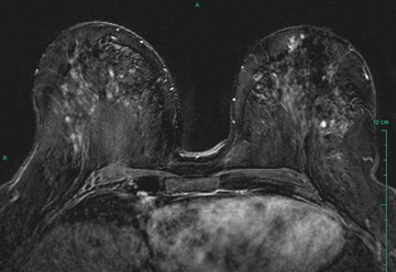 Figure 1. An MRI image of the breast showing a false positive nodule. High-res (TIF) version (Right-click and Save As) |
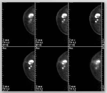 Figure 2. Images of the breast taken using PEM. PEM images depict additional small lesions missed by a whole body PET scan. High-res (TIF) version (Right-click and Save As) |
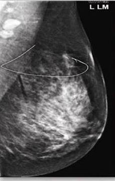 Figure 3. Mammography image of a left breast showing a lesion. High-res (TIF) version (Right-click and Save As) |
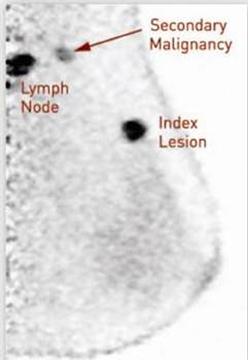 Figure 4. Image of a left breast taken using PEM. PEM depicts an index lesion and additional lesion missed by mammography. High-res (TIF) version (Right-click and Save As) |
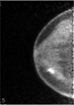 Figure 5. Image of a right breast with an irregular mass taken using PEM. High-res (TIF) version (Right-click and Save As) |
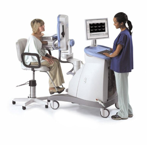 Figure 6. Depiction of a PEM Scanner. High-res (TIF) version (Right-click and Save As) |
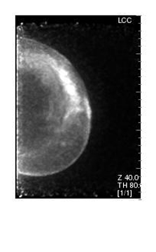 Figure 7. Depiction of a PEM image illustrating a breast implant and cancer. High-res (TIF) version (Right-click and Save As) |
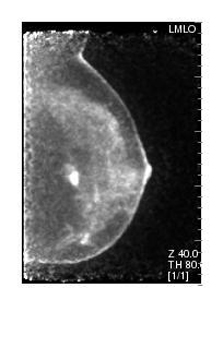 Figure 8. Depiction of a PEM image illustrating a breast implant and cancer. High-res (TIF) version (Right-click and Save As) |
 Figure 9. Depiction of a PEM image illustrating a breast implant and incidental cancer. High-res (TIF) version (Right-click and Save As) |
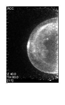 Figure 10. Depiction of a PEM image illustrating a breast implant and incidental cancer. High-res (TIF) version (Right-click and Save As) |
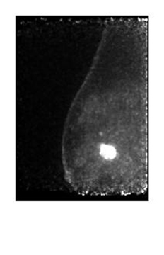 Figure 11. Depiction of a PEM image illustrating invasive cancer within the breast at the biopsy site. High-res (TIF) version (Right-click and Save As) |
 Figure 12. Depiction of a PEM image showing invasive cancer within the breast. High-res (TIF) version (Right-click and Save As) |
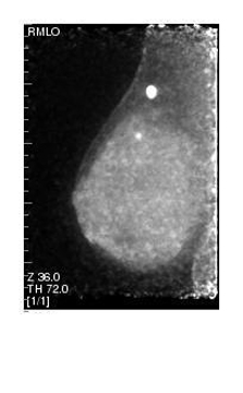 Figure 13. PEM image showing multifocal cancer within the breast. High-res (TIF) version (Right-click and Save As) |
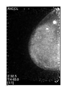 Figure 14. PEM image showing multifocal cancer within the breast. High-res (TIF) version (Right-click and Save As) |
|
|
 PDF
PDF