RSNA Press Release
- A 12-year study has found that laser ablation with MR guidance has comparable results to open surgery in the treatment of liver tumors.
- Laser ablation is a minimally invasive procedure that uses laser light to destroy tumors.
- Laser ablation has lower rates of morbidity and mortality than surgery.
- Laser ablation can be used to treat multiple tumors simultaneously.
MR-Guided Laser Effective in Treating Liver Tumors
Released: November 29, 2005
| Media Contacts: | |
| RSNA Media Relations: | (630) 590-7762 |
| Maureen Morley (630) 590-7754 mmorley@rsna.org |
Heather Babiar (630) 590-7738 hbabiar@rsna.org |
CHICAGO - A large-scale, 12-year study has found that laser ablation with magnetic resonance (MR) guidance is as effective as traditional surgery in the treatment of liver tumors in some patients. The study was presented today at the annual meeting of the Radiological Society of North America (RSNA).
In the largest study of its type with the longest follow-up, 839 patients at the University of Frankfurt in Germany received MR-guided laser-induced thermotherapy (LITT) for the treatment of liver tumors resulting from colorectal cancer. Between 1993 and 2005, the researchers treated 2,506 liver tumors and tracked survival rates to evaluate the long-term results of the procedure. The average survival rate from the date of diagnosis was 3.8 years, which compares favorably to survival rates after traditional surgery (approximately 1.5 to 5.0 years).
In LITT, also known as laser ablation, laser light is used to destroy tumor tissue. According to the study's lead author, Martin Mack, M.D., laser ablation has many advantages over other treatment methods.
"Traditional surgical resection has higher morbidity and mortality rates than laser ablation," said Dr. Mack, an associate professor in the department of diagnostic and interventional radiology at the University of Frankfurt. "Laser treatment can be done on an outpatient basis under local anesthesia. Typically, the patient stays only a couple of hours, instead of a couple of weeks in the hospital after surgical liver resection," he said.
Laser ablation can be used to treat tumors that occur in both halves of the liver—often during the same treatment—which is practically impossible in a traditional surgery where typically only the left or right lobe is resected. If new tumors are found during follow-up exams, it is much easier to repeat laser treatment than to subject the patient to another open surgery.
Dr. Mack believes that laser combined with MR guidance will have wide-ranging impact on the treatment of tumors throughout the body, and may one day replace traditional surgery as the gold standard of treatment.
"Many surgeons are already performing local ablation instead of resection, because they have already recognized the positive effect of local ablation," he said. "I believe that minimally invasive tumor ablation together with chemotherapy will play the most important role in the treatment of tumors in the years to come."
Co-authors are Katrin Eichler, M.D., Thomas Lehnert, M.D., Dirk Proschek, M.D., Joern O. Balzer, M.D., and Thomas J. Vogl, M.D.
# # #
Note: Copies of RSNA 2005 news releases and electronic images will be available online at RSNA.org/press05 beginning Monday, Nov. 28.
RSNA is an association of more than 38,000 radiologists, radiation oncologists, medical physicists and related scientists committed to promoting excellence in radiology through education and by fostering research, with the ultimate goal of improving patient care. The Society is based in Oak Brook, Ill.
Editor's note: The data in these releases may differ from those in the printed abstract and those actually presented at the meeting, as researchers continue to update their data right up until the meeting. To ensure you are using the most up-to-date information, please call the RSNA Newsroom at (312) 949-3233.
| Abstract: |
Images (.JPG format)
 Figure 1. Close up of a laser used in minimally invasive laser therapy. |
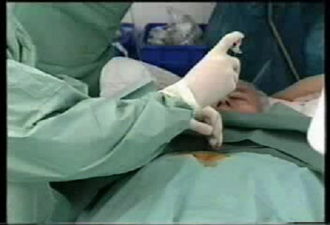 Figure 2. Interventional radiologist performing laser therapy. |
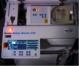 Figure 3. Photograph of the machine used in laser therapy. |
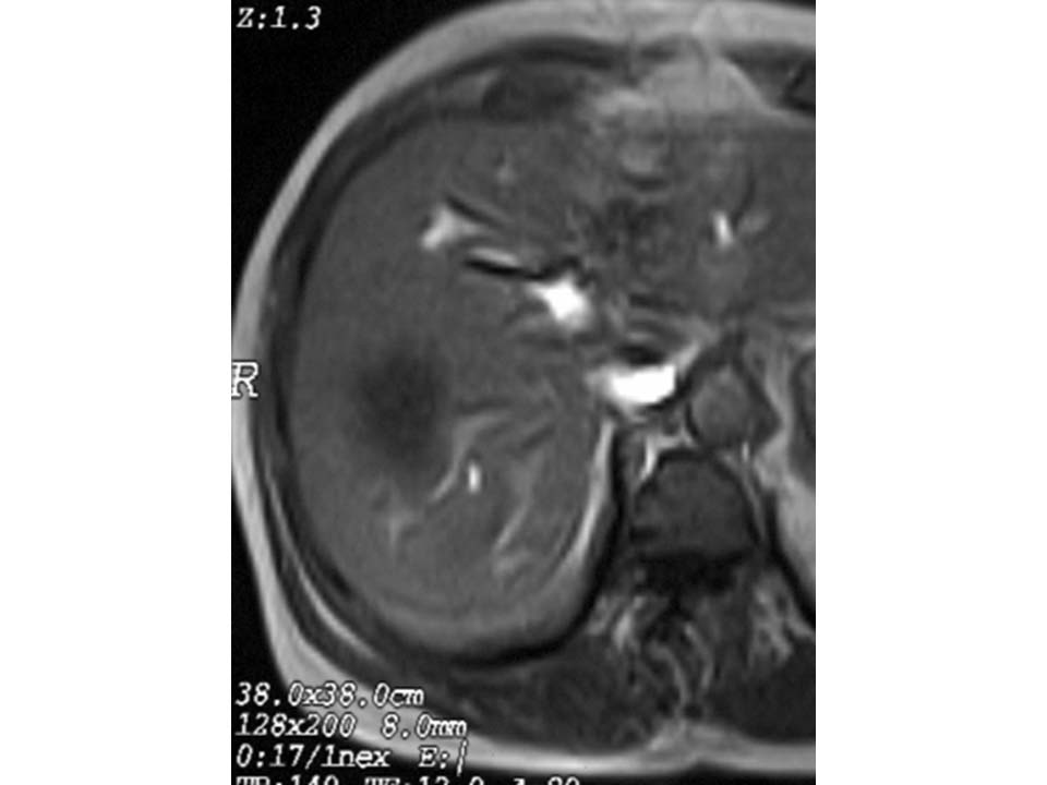 Figure 4.1. Liver metastases before LITT High-res (TIF) version (Right-click and Save As) |
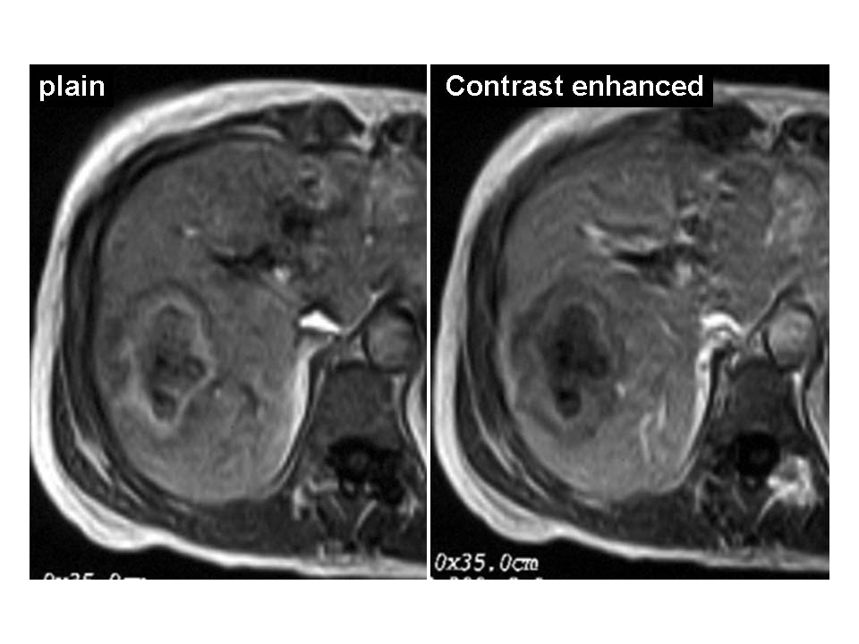 Figure 4.2. Liver metastases 24 hours after LITT (left image non contrast enhanced, right image after administration of a contrast agent. High-res (TIF) version (Right-click and Save As) |
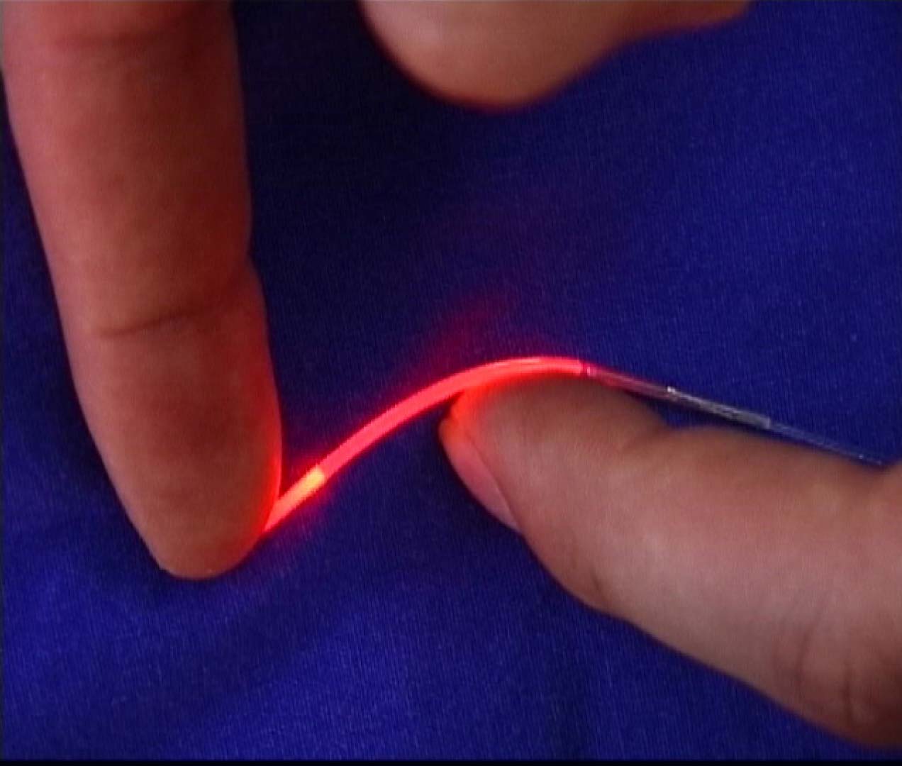 Figure 4.3. High-res (TIF) version (Right-click and Save As) |
 Figure 4.4. High-res (TIF) version (Right-click and Save As) |
 Figure 4.5. High-res (TIF) version (Right-click and Save As) |
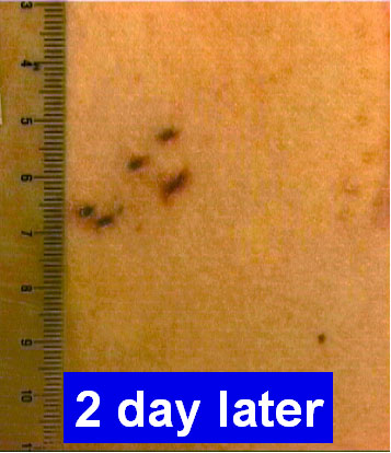 Figure 5. Close up photograph of patient's skin two days after she received laser treatment. |
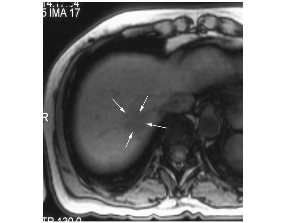 Figure 6.1 Liver metastases before LITT, non contrast enhanced image High-res (TIF) version (Right-click and Save As) |
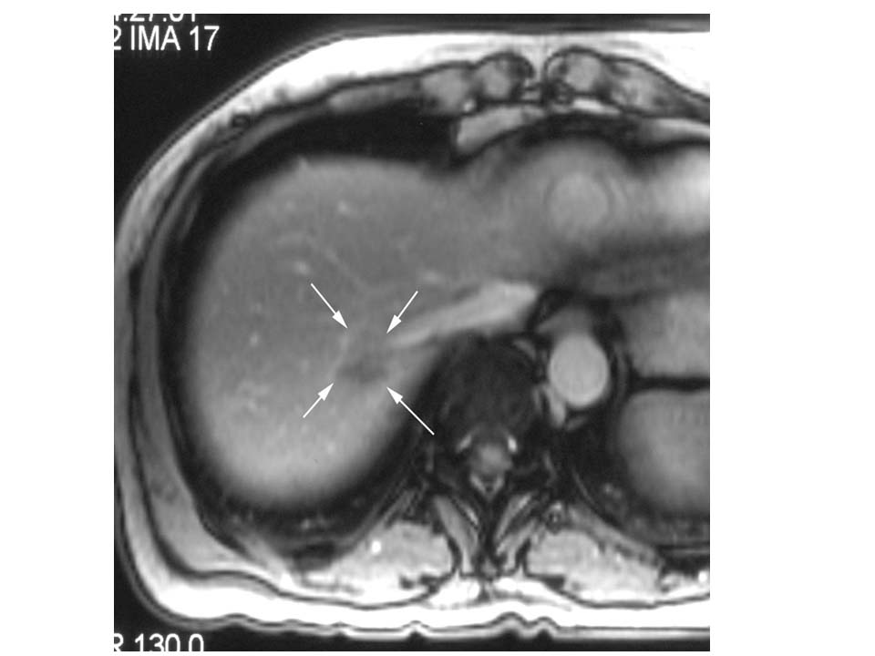 Figure 6.2 Liver metastases before LITT, contrast enhanced image High-res (TIF) version (Right-click and Save As) |
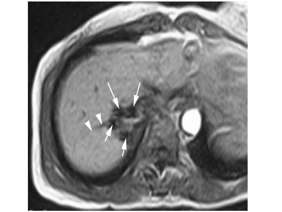 Figure 6.3 This image demonstrates the placement of the laser applicators (arrows) in axial slice orientation, arrow heads are showing a liver vein. High-res (TIF) version (Right-click and Save As) |
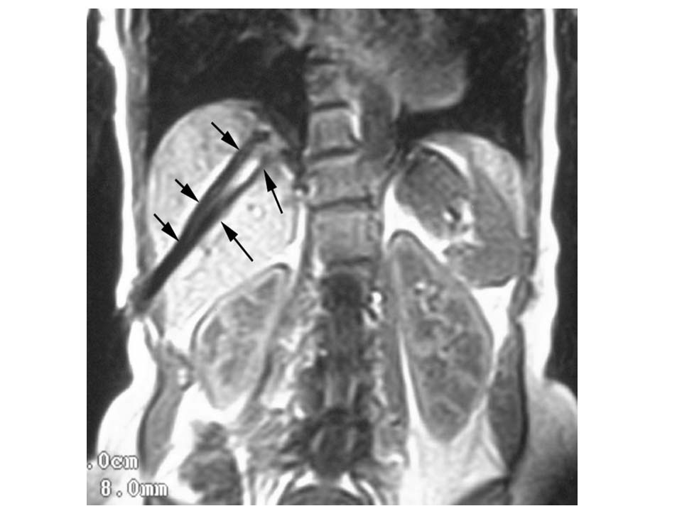 Figure 6.4 This image demonstrates the placement of the laser applicators (arrows) in coronal slice orientation High-res (TIF) version (Right-click and Save As) |
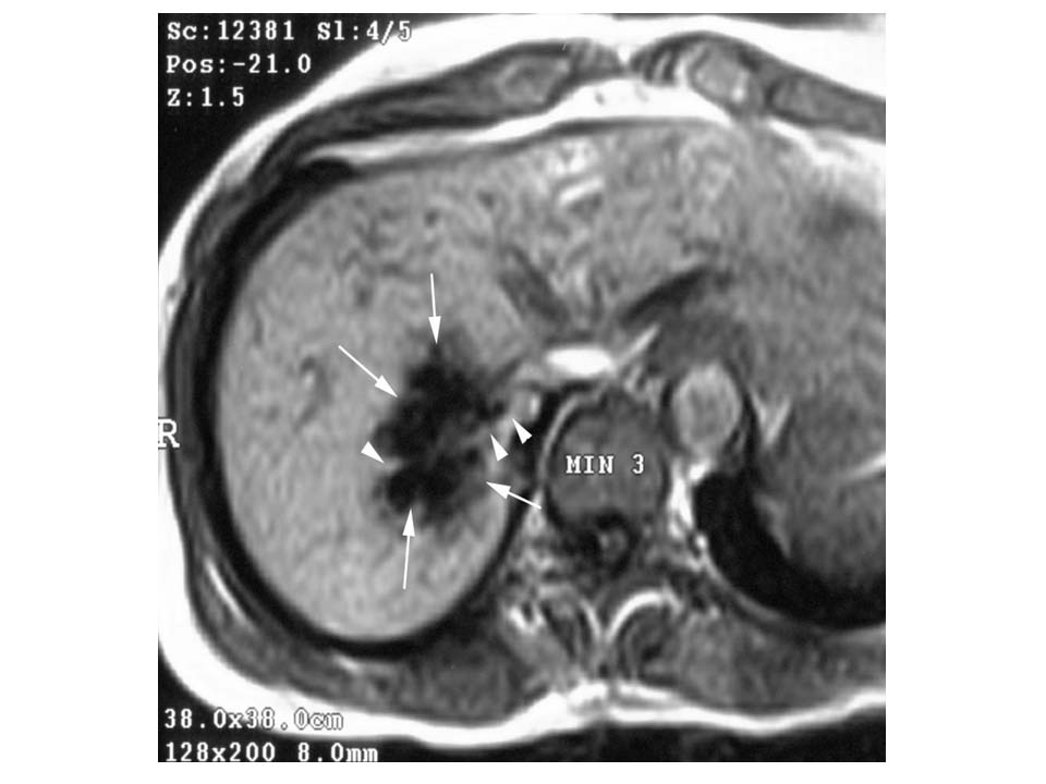 Figure 6.5 The image shows the thermal effect after 3 minutes of LITT (decrease in signal intensity due to heating of the tissue up to 100°C High-res (TIF) version (Right-click and Save As) |
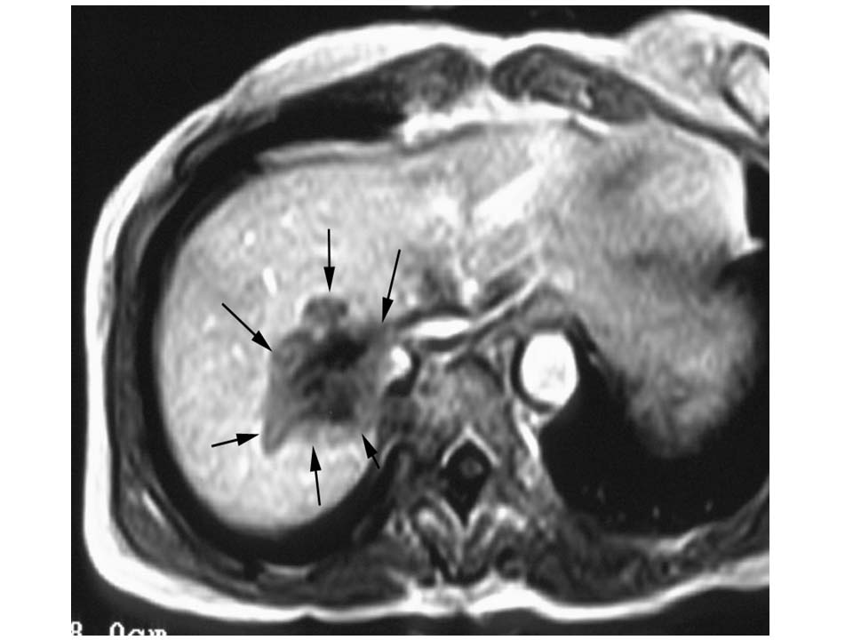 Figure 6.6 The contrast enhanced image shows the induced coagulation necrosis immediately after LITT High-res (TIF) version (Right-click and Save As) |
 PDF
PDF