RSNA Press Release
- Virtual colonoscopy with CAD is a 15-minute procedure requiring no patient sedation that can substitute for conventional fiberoptic colonoscopy for selected patients.
- Computer-aided detection (CAD) technology can improve the performance of virtual colonoscopy in colon cancer screening.
- CAD marks abnormalities on virtual colonoscopy images for review by the radiologist.
Virtual Colonoscopy Performance Enhanced by Computer-Aided Detection
Released: November 28, 2005
| Media Contacts: | |
| RSNA Media Relations: | (630) 590-7762 |
| Maureen Morley (630) 590-7754 mmorley@rsna.org |
Heather Babiar (630) 590-7738 hbabiar@rsna.org |
CHICAGO - Computed tomography (CT) colonography with computer-aided detection (CAD) is highly effective for finding colon polyps, according to a large-scale, multi-center study conducted by the National Institutes of Health (NIH) and presented today at the annual meeting of the Radiological Society of North America (RSNA).
CT colonography, commonly called virtual colonoscopy, is a minimally invasive exam that physicians hope will encourage more people to be screened for colon cancer. Virtual colonoscopy is desirable because there is no risk of bleeding or colon perforation and intravenous sedation is unnecessary. The procedure is less costly than conventional colonoscopy and more convenient, taking 15 minutes or less.
"The performance of virtual colonoscopy continues to improve, and the exam will become a colorectal cancer screening method more patients and doctors will find acceptable," said the study's senior investigator Ronald M. Summers, M.D., Ph.D. Dr. Summers is staff radiologist, chief of the clinical image processing service and chief of the virtual endoscopy and computer-aided diagnosis laboratory at the NIH clinical center in Bethesda, Md.
Dr. Summers and colleagues studied 792 patients at three medical centers using virtual colonoscopy with CAD to detect adenomatous colon polyps eight millimeters (mm) and larger. Colon polyps are benign growths that may develop into colon cancer if not removed.
CT colonography produces 600 to 1,000 images per patient. Dr. Summers likened the interpretation process to the "needle in a haystack problem." With CAD technology, after the radiologist has interpreted the images, the computer acts as a second set of eyes, reviewing the images and marking abnormalities for the radiologist to review. CAD has the potential to find polyps that a radiologist might miss.
"We think that CAD will help improve the performance of virtual colonoscopy by reducing the perceptual error that can occur when radiologists have to read the large number of images in these studies," Dr. Summers said.
With CAD the researchers detected polyps in 89.3 percent of patients with polyps 10 mm or larger and 85.4 percent of patients with polyps 8 mm or larger. CAD's false positive rates were 2.1 per patient for polyps 10 mm and larger and 6.7 per patient for polyps 8 mm and larger, which fall within the researchers' acceptable limits.
Dr. Summers predicted that soon all physicians who are interpreting virtual colonoscopy will want to have CAD readily at hand. "I think CAD is soon to become a mainstream technology," he said.
The American Cancer Society recommends that people at average risk for colon cancer begin regular colon cancer screening at age 50, but current compliance with this recommendation remains well below 50 percent.
Co-authors are Jack Yao, Ph.D., Perry Pickhardt, M.D., Marek Franaszek, Ph.D., Ingmar Bitter, Ph.D., Jong-Ho Choi, Sc.D., M.D., Daniel Brickman and Vamsi Krishna.
A related study on this topic entitled CAD in Minimal-Preparation CT Colonography: Technical Feasibility will be presented by Janne J. Nappi, Ph.D., Wednesday, Nov. 30 at 11:20 a.m. in Room S401CD.
# # #
Note: Copies of RSNA 2005 news releases and electronic images will be available online at RSNA.org/press05 beginning Monday, Nov. 28.
RSNA is an association of more than 38,000 radiologists, radiation oncologists, medical physicists and related scientists committed to promoting excellence in radiology through education and by fostering research, with the ultimate goal of improving patient care. The Society is based in Oak Brook, Ill.
Editor's note: The data in these releases may differ from those in the printed abstract and those actually presented at the meeting, as researchers continue to update their data right up until the meeting. To ensure you are using the most up-to-date information, please call the RSNA Newsroom at (312) 949-3233.
| Abstract: |
Images (.JPG format)
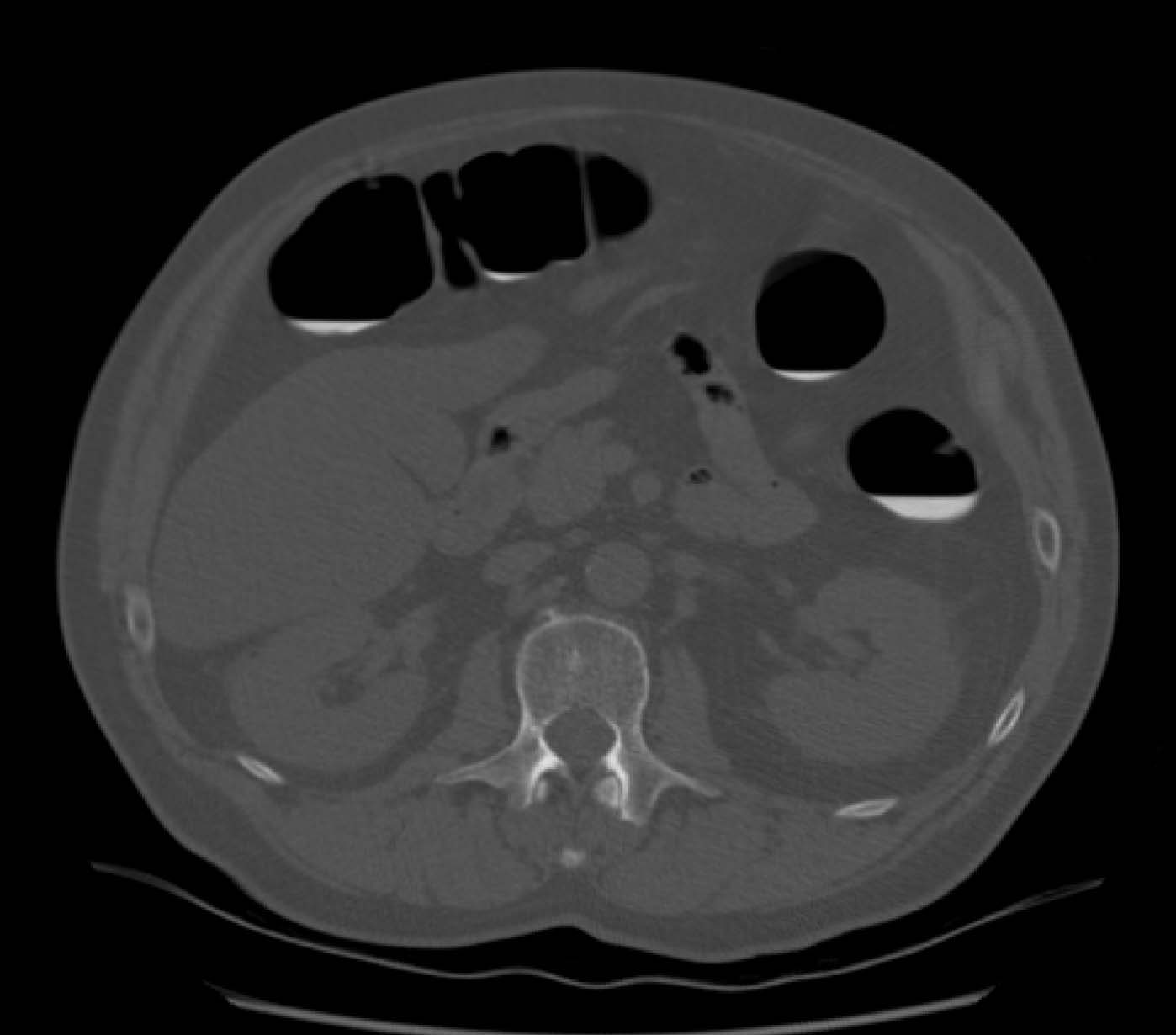 Figure 1. Virtual colonoscopy technique – air insufflation, supine and prone MDCT and single breathhold. High-res (TIF) version (Right-click and Save As) |
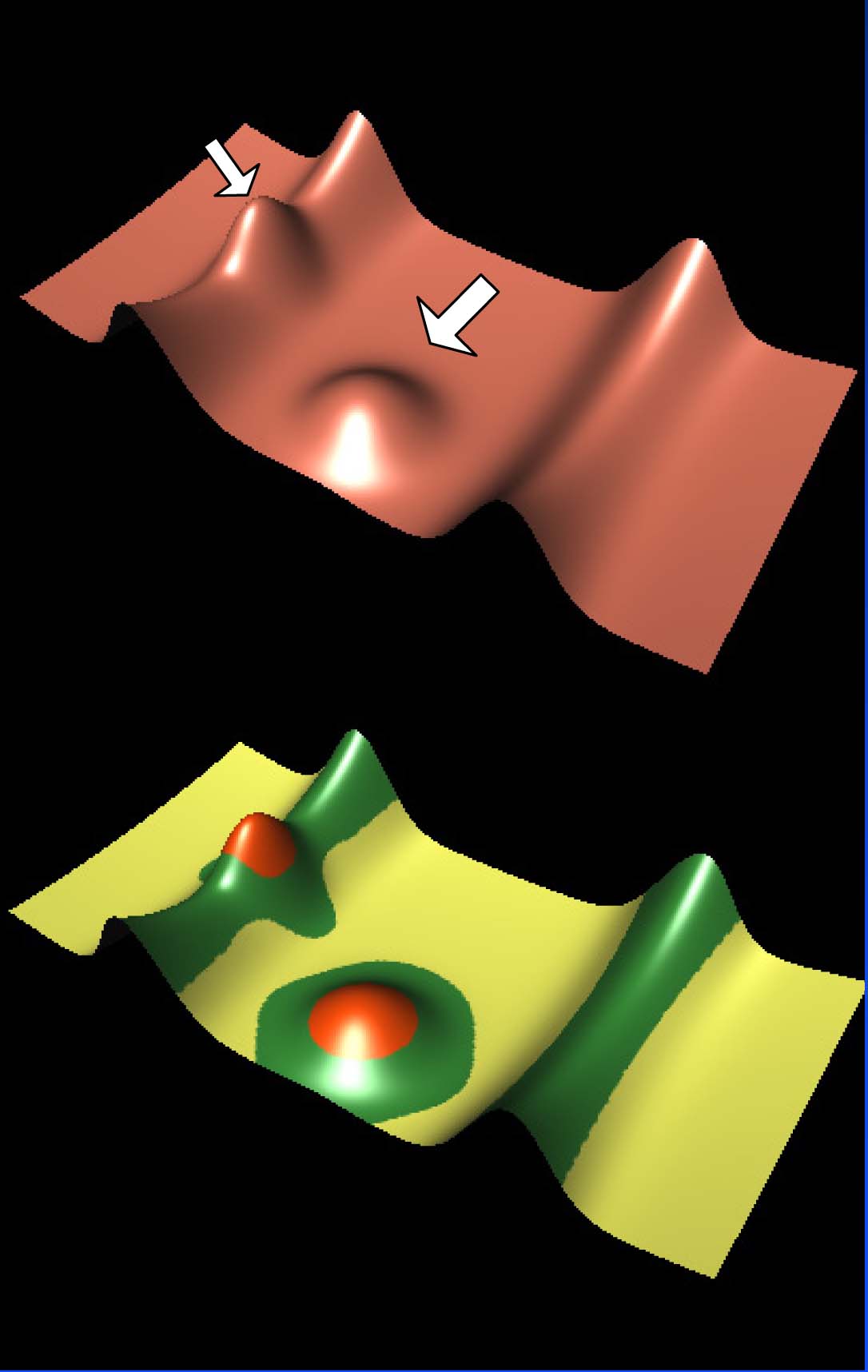 Figure 2. Multi-stage analysis using CAD system – locate air- and fluid-filled parts of colon, analyze surface shape of colon, locate entire 3-D borders of polyp candidates and distinguish true and false positive polyp candidates. High-res (TIF) version (Right-click and Save As) |
 Figure 3. Illustration of various colons. High-res (TIF) version (Right-click and Save As) |
| Figure 4. — A photograph and two illustrations of a 1.4 cm transverse colon polyp in 64-year-old woman. | ||
 Figure 4.1. A photograph of a 1.4 cm transverse colon polyp in 64-year-old woman. High-res (TIF) version (Right-click and Save As) |
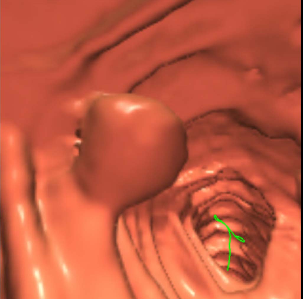 Figure 4.2. An illustration of a 1.4 cm transverse colon polyp in 64-year-old woman. High-res (TIF) version (Right-click and Save As) |
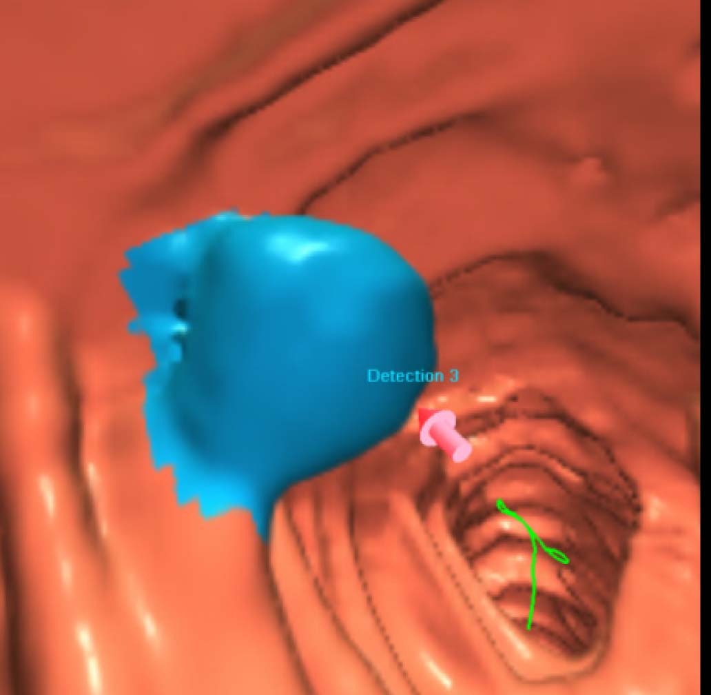 Figure 4.3. An illustration of a 1.4 cm transverse colon polyp in 64-year-old woman. High-res (TIF) version (Right-click and Save As) |
| Figure 5. — A photograph an two illustrations of an 0.8 cm sigmoid colon polyp in a 60-year-old man. | ||
 Figure 5.1. A photograph an 0.8 cm sigmoid colon polyp in a 60-year-old man. High-res (TIF) version (Right-click and Save As) |
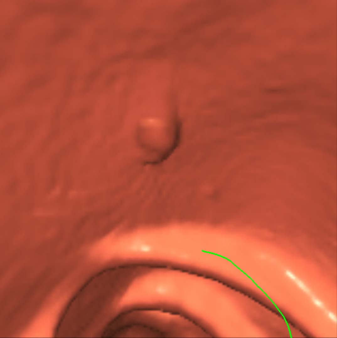 Figure 5.2. An illustration an 0.8 cm sigmoid colon polyp in a 60-year-old man. High-res (TIF) version (Right-click and Save As) |
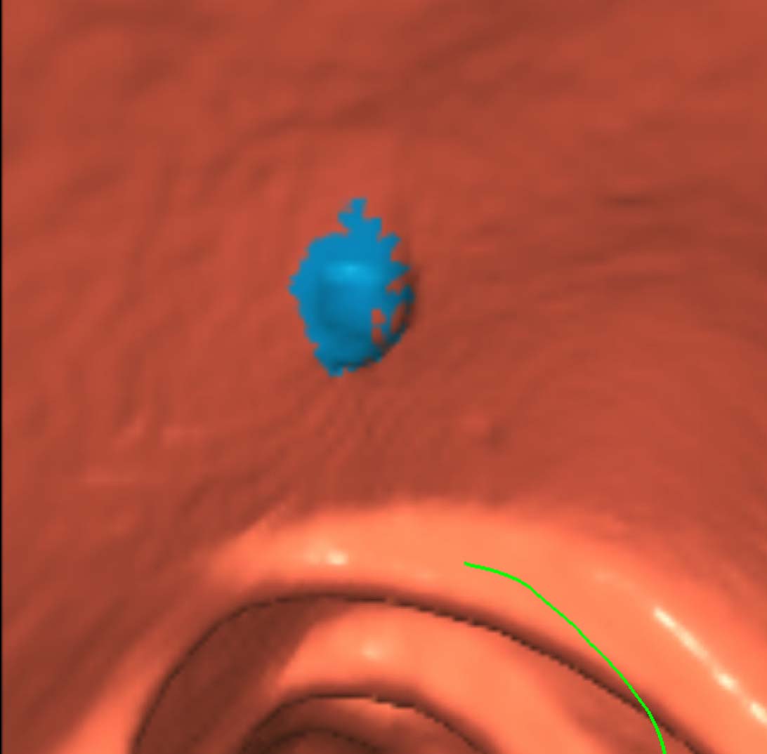 Figure 5.3. An illustration an 0.8 cm sigmoid colon polyp in a 60-year-old man. High-res (TIF) version (Right-click and Save As) |
| Figure 6. — A photograph and two illustrations of a 0.6 cm transverse colon polyp in a 65-year-old man. | ||
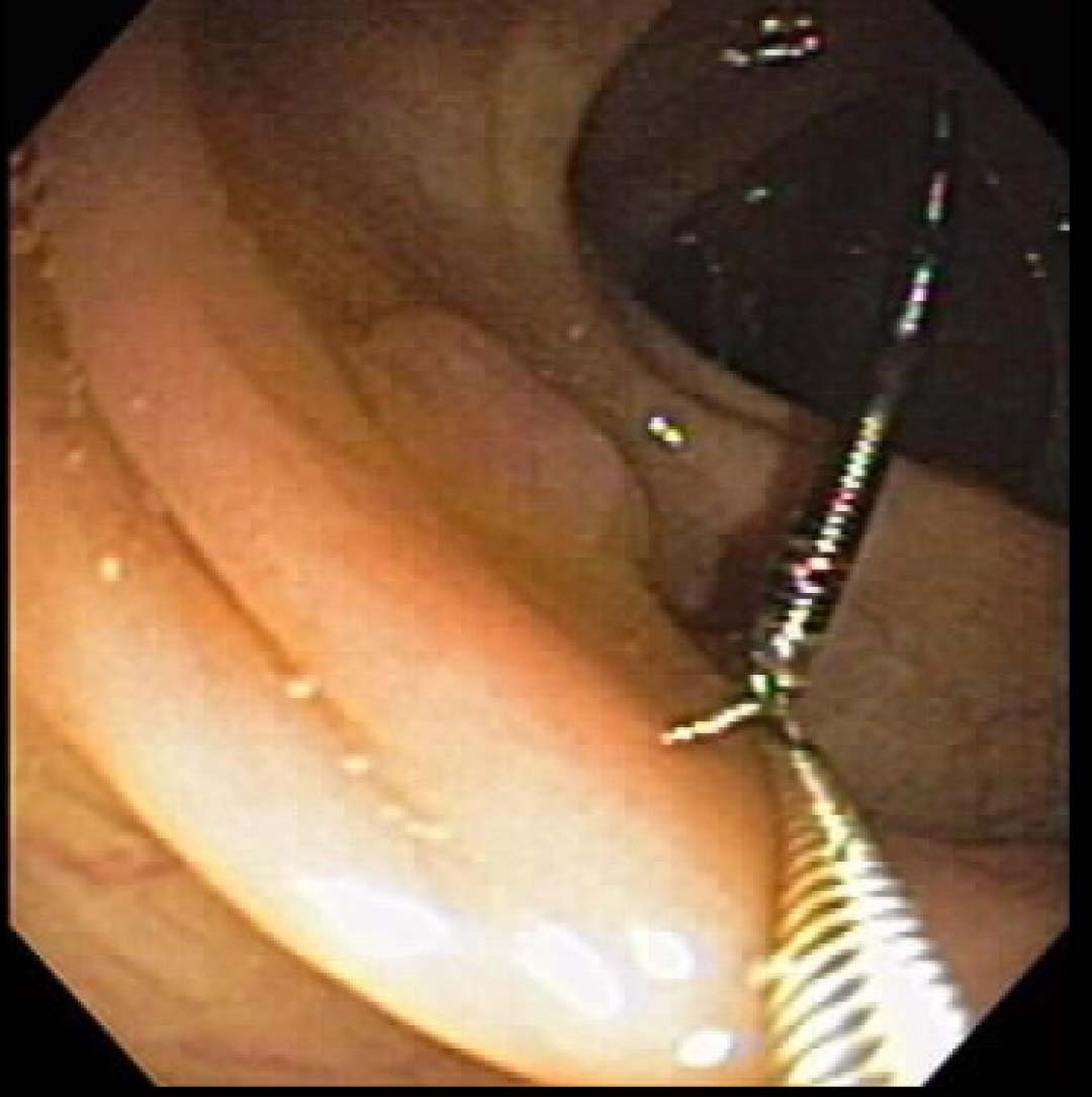 Figure 6.1. A photograph of a 0.6 cm transverse colon polyp in a 65-year-old man. High-res (TIF) version (Right-click and Save As) |
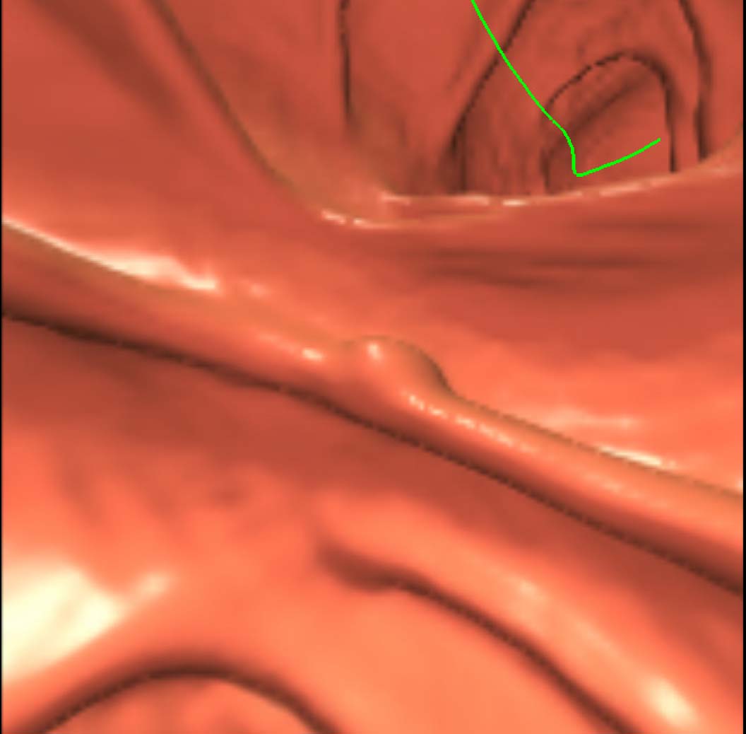 Figure 6.2. An illustration of a 0.6 cm transverse colon polyp in a 65-year-old man. High-res (TIF) version (Right-click and Save As) |
 Figure 6.3. An illustration of a 0.6 cm transverse colon polyp in a 65-year-old man. High-res (TIF) version (Right-click and Save As) |
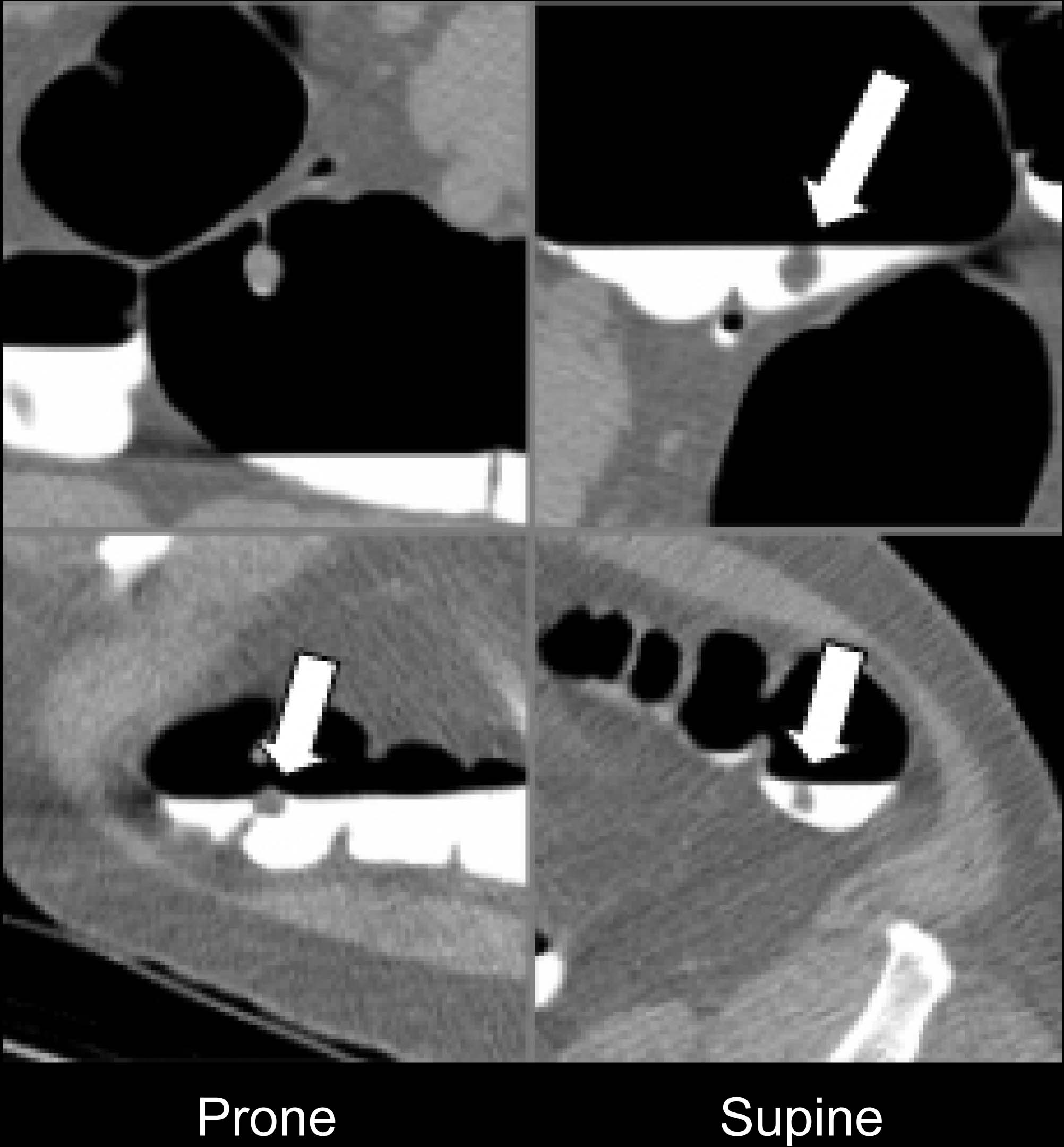 Figure 7. Illustration of false negatives – air-fluid boundary and on or adjacent to haustral folds. High-res (TIF) version (Right-click and Save As) |
 Figure 8. Illustration of false positives – ileocecal valves and haustral folds. High-res (TIF) version (Right-click and Save As) |
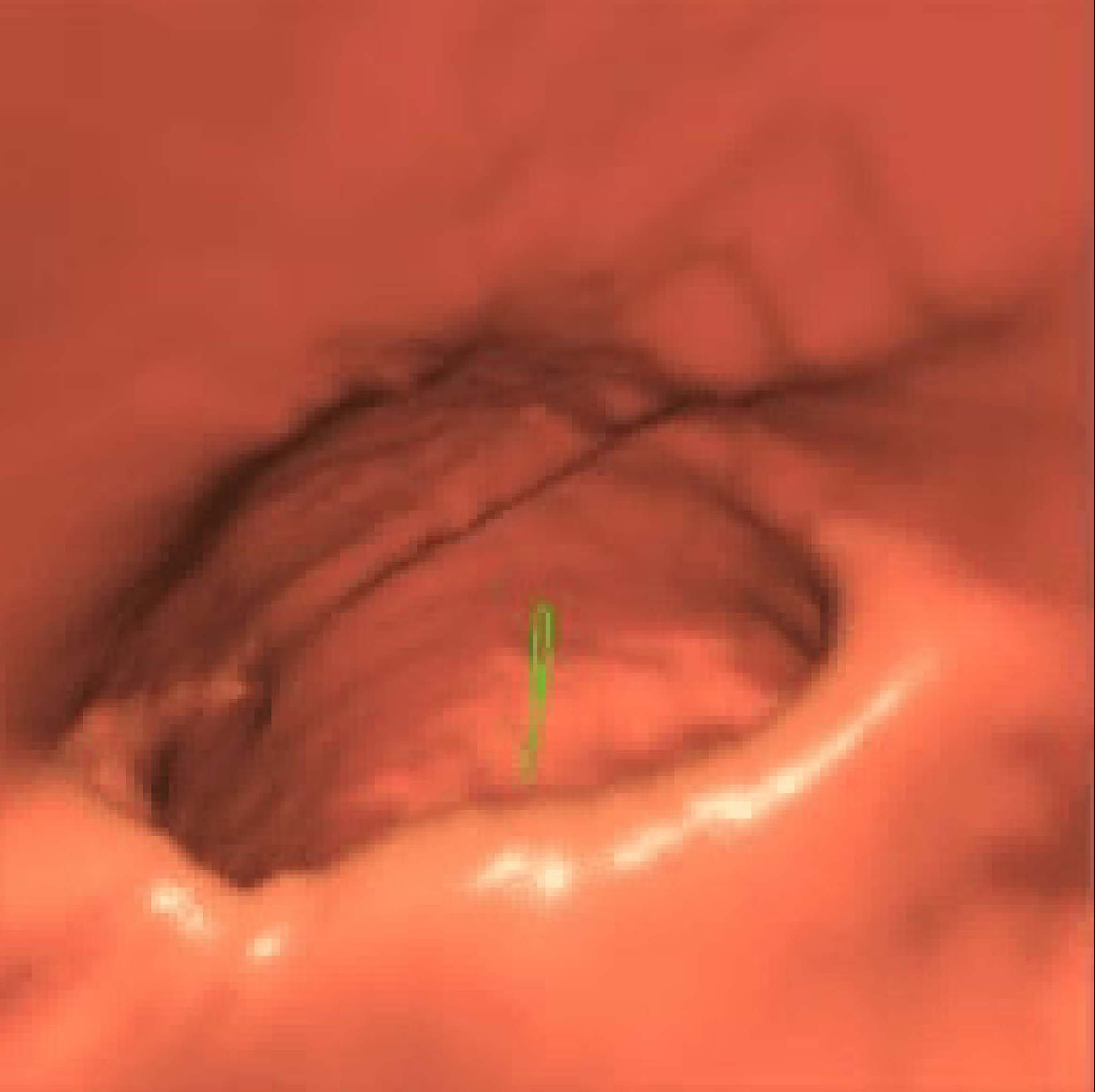 Figure 9. CAD illustration. High-res (TIF) version (Right-click and Save As) |
 PDF
PDF