Researchers Use Gaming Technology to Create Better X-Rays
Released: December 01, 2015
At A Glance
- Software developed for Microsoft Kinect can automatically measure body thickness, and check for motion and positioning prior to X-ray imaging.
- Through real-time monitoring, the software alerts the user to factors that could compromise image quality.
- Patients will benefit from reduced radiation exposure and higher quality images to ensure diagnostic accuracy.
- RSNA Media Relations
1-630-590-7762
media@rsna.org - Maureen Morley
1-630-590-7754
mmorley@rsna.org - Linda Brooks
1-630-590-7738
lbrooks@rsna.org
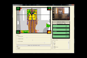
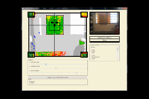
CHICAGO — Researchers have developed software for the Microsoft Kinect gaming console that measures body part thickness and checks for motion, positioning and beam adjustment immediately before X-ray imaging, according to a feasibility study presented today at the annual meeting of the Radiological Society of North America (RSNA).

"Patients, technologists and radiologists want the best quality X-rays at the lowest dose possible without repeating images," said Steven Don, M.D., associate professor of radiology at Mallinckrodt Institute of Radiology, Washington University School of Medicine in St. Louis, Mo. "This technology is a tool to help achieve that goal. Patients will benefit from reduced radiation exposure and higher quality images to ensure diagnostic accuracy."
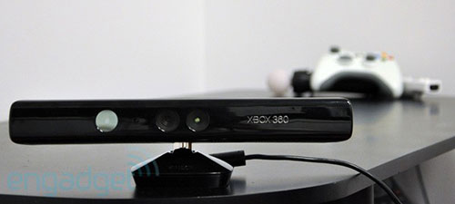
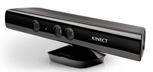
Microsoft Kinect was originally developed as a motion sensor and facial and voice recognition device for the Xbox gaming system that enabled players to play games without a standard controller. Subsequently, the technology has been adapted for select non-gaming applications.
For this feasibility study, Dr. Don and colleagues combined the technology of the Microsoft Kinect 1.0 with proprietary software to address common problems that affect imaging results, including body-part thickness and motion.
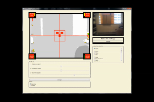
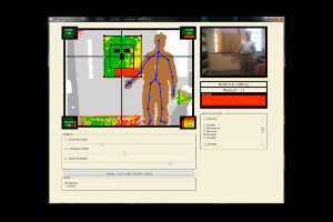
"To optimize radiation exposure and image quality, X-ray technique should be set based on body-part thickness," Dr. Don explained. "Use of traditional calipers is time-consuming, intrusive, and sometimes frightening to young children. Using Microsoft Kinect with this software, we can measure body-part thickness automatically without patient contact."
Additionally, the software provides valuable information on motion and positioning with respect to automatic exposure control (AEC) sensors, image receptor and body part within the X-ray field. Through real-time monitoring, the software alerts the user when any of these factors do not match the requisition—such as the wrong body part—or could compromise image quality. This fail-safe helps to reduce or eliminate common causes of unnecessary repeat image acquisition.
"This device can help technologists and radiologists achieve the radiation dose goal of ALARA, As Low As Reasonably Achievable, while enhancing the quality and consistency of X-ray images," Dr. Don said.
Dr. Don noted that the radiology research community is constantly improving imaging for patients to ensure accurate diagnoses while striving to reduce dose.
"In the future, we hope to see this device, and other tools like it, installed on radiography equipment to aid technologists by identifying potential problems before they occur," he said.
Co-authors on the study are Robert MacDougall, M.Sc., and William Clayton.
Note: Copies of RSNA 2015 news releases and electronic images will be available online at RSNA.org/press15 beginning Monday, Nov. 30.
RSNA is an association of more than 54,000 radiologists, radiation oncologists, medical physicists and related scientists, promoting excellence in patient care and health care delivery through education, research and technologic innovation. The Society is based in Oak Brook, Ill. (RSNA.org)
Editor's note: The data in these releases may differ from those in the published abstract and those actually presented at the meeting, as researchers continue to update their data right up until the meeting. To ensure you are using the most up-to-date information, please call the RSNA Newsroom at 1-312-791-6610.
For patient-friendly information on X-ray imaging, visit RadiologyInfo.org.
Images (.JPG and .TIF format)
Videos clips (.mp4 format)
Video 2. Researchers setting up the equipment for left wrist exam.
Download.mp4
(Right-click and Save As)
Video 3. Researchers are testing body movement on the monitor.
Download.mp4
(Right-click and Save As)
Video 4. Researchers program the equipment for a left elbow exam. The red box on the display alerts the technician that the wrong elbow is being set up for imaging.
Download.mp4
(Right-click and Save As)