RSNA Press Release
- Engaging in light exercise may protect against cartilage degeneration and osteoarthritis of the knee.
- Women at risk for osteoarthritis who engage in moderate to strenuous exercise may accelerate damage to the knee cartilage.
- Frequent knee-bending activities are also associated with increased risk of osteoarthritis.
- More than 27 million American adults have osteoarthritis.
Light Exercise May Prevent Osteoarthritis
Released: November 29, 2010
| Media Contacts: | RSNA Newsroom | 1-312-949-3233 |
| Before 11/27/2010 or after 12/02/2010: | RSNA Media Relations: | 1-630- 590-7762 |
| |
Linda Brooks 1-630-590-7738 lbrooks@rsna.org |
Maureen Morley 1-630-590-7754 mmorley@rsna.org |
CHICAGO — People at risk for osteoarthritis may be able to delay the onset of the disease or even prevent it with simple changes to their physical activity, according to a study presented today at the annual meeting of the Radiological Society of North America (RSNA).
"According to the results of our study, participating in a high-impact activity, such as running, more than one hour per day at least three times a week appears associated with more degenerated cartilage and potentially a higher risk for development of osteoarthritis," said the study's senior author Thomas M. Link, M.D., professor of radiology and chief of musculoskeletal imaging at the University of California, San Francisco (UCSF). "On the other hand, engaging in light exercise and refraining from frequent knee-bending activities may protect against the onset of the disease."
Osteoarthritis is a degenerative joint disease that causes pain, swelling and stiffness. According to the National Institute of Arthritis and Musculoskeletal and Skin Diseases, osteoarthritis is the most common form of arthritis and affects an estimated 27 million Americans over the age of 25.
For the study, the researchers recruited 132 asymptomatic participants at risk for knee osteoarthritis who were enrolled in the National Institutes of Health Osteoarthritis Initiative, as well as 33 age- and body mass index-matched controls. Study participants included 99 women and 66 men between the ages of 45 and 55. The participants were separated into three exercise and strength-training levels, based on their responses to the Physical Activity Scale for the Elderly (PASE) questionnaire. Exercise levels included sedentary, light exercisers and moderate to strenuous exercisers, strength-training groups included none, minimal and frequent. Knee-bending activities were also analyzed.
MRI exams revealed that light exercisers had the healthiest knee cartilage among all exercise levels, and patients with minimal strength training had healthier cartilage than patients with either no strength training or frequent strength training.
Moderate to strenuous exercise in women who did any amount of strength training was associated with higher water content and more degenerated collagen architecture in the knee.
"The results for this group indicate that moderate to strenuous exercise may accelerate cartilage degeneration, putting these women at even greater risk of developing osteoarthritis," said study coauthor Keegan K. Hovis, B.S., R.N., research associate in the Department of Radiology at UCSF.
In addition, the findings showed that frequent knee-bending activities, such as climbing up at least 10 flights of stairs a day, lifting objects weighing more than 25 pounds, or squatting, kneeling or deep knee bending for at least 30 minutes per day, were associated with higher water content and cartilage abnormalities.
According to Dr. Link, known risk factors for cartilage degeneration include excess weight, knee injuries, frequent knee bending and severe or strenuous physical activity.
"People can reduce their risk for osteoarthritis by maintaining a healthy weight and avoiding risky activities and strenuous exercise," he said. "Lower-impact sports, such as walking, swimming or using an elliptical trainer are likely more beneficial than high-impact sports, such as running or tennis."
"Our findings indicate that light exercise, particularly frequent walking, is a safer choice in maintaining healthy cartilage," Hovis added.
Other coauthors are Christoph Stehling, M.D., Jean-Baptiste Pialat, M.D., Michael C. Nevitt, Ph.D., M.P.H., and Charles E. McCulloch, Ph.D.
# # #
Note: Copies of RSNA 2010 news releases and electronic images will be available online at RSNA.org/press10 beginning Monday, Nov. 29.
RSNA is an association of more than 44,000 radiologists, radiation oncologists, medical physicists and related scientists committed to excellence in patient care through education and research. The Society is based in Oak Brook, Ill. (RSNA.org)
Editor's note: The data in these releases may differ from those in the printed abstract and those actually presented at the meeting, as researchers continue to update their data right up until the meeting. To ensure you are using the most up-to-date information, please call the RSNA Newsroom at 1-312-949-3233.
For patient-friendly information on MRI, visit RadiologyInfo.org.
| Abstract: |
Presenter Interview:
- Video clip
Thomas M. Link, M.D., sound bite - Video clip
Thomas M. Link, M.D., sound bite
Images (.JPG format)
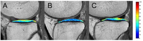
Figure 1: This figure shows three color-coded T2 maps overlaid on first-echo MR images that show (A) high lateral tibia T2 values in a sedentary individual; (B) low lateral tibia T2 values in a light exerciser; (C) high lateral tibia T2 values in a moderate-strenuous exerciser who had risk factors for osteoarthritis. High-res (TIF) version (Right-click and Save As) |
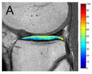
Figure 1a: This figure shows a three-color coded T2 map overlaid on a first-echo MR image, showing high lateral tibia T2 values in a sedentary individual. High-res (TIF) version (Right-click and Save As) |
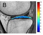
Figure 1b: This figure shows a three-color coded T2 map overlaid on a first-echo MR image, showing low lateral tibia T2 values in a light exercise. High-res (TIF) version (Right-click and Save As) |
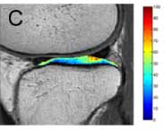
Figure 1c: This figure shows a three-color coded T2 map overlaid on a first-echo MR image, showing high lateral tibia T2 values in a moderate-strenuous exerciser who had risk factors for osteoarthritis. High-res (TIF) version (Right-click and Save As) |
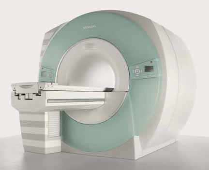
Trio 1: A photograph of the Siemens Trio MRI System. High-res (TIF) version (Right-click and Save As) |

Trio 2: A photograph of the Siemens Trio MRI System. High-res (TIF) version (Right-click and Save As) |
 PDF
PDF