Researchers Use 3-D Printing to Guide Human Face Transplants
Released: December 01, 2014
At A Glance
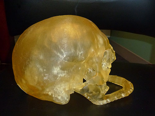
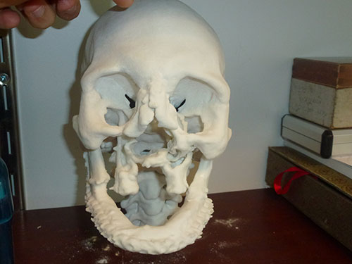
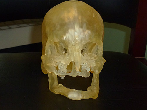
- Life-size custom skull reproductions made using CT images and a 3-D printer help surgical teams plan and perform face transplantation procedures.
- The 3-D printed models provide superior pre-operative data and reduce total procedure time.
- Based on the results of this study, 3-D printing is now a mandatory step in surgical planning for face transplantation procedures at Brigham and Women’s Hospital.
- RSNA Media Relations
1-630-590-7762
media@rsna.org - Maureen Morley
1-630-590-7754
mmorley@rsna.org - Linda Brooks
1-630-590-7738
lbrooks@rsna.org
Chicago — Researchers are using computed tomography (CT) and 3-D printing technology to recreate life-size models of patients' heads to assist in face transplantation surgery, according to a study presented today at the annual meeting of the Radiological Society of North America (RSNA).
Physicians at Brigham and Women's Hospital in Boston performed the country's first full-face transplantation in 2011 and have subsequently completed four additional face transplants. The procedure is performed on patients who have lost some or all of their face as a result of injury or disease.
In the study, a research team led by Frank J. Rybicki, M.D., radiologist and director of the hospital's Applied Imaging Science Laboratory, Bohdan Pomahac, M.D., lead face transplantation surgeon, and Amir Imanzadeh, M.D., research fellow, assessed the clinical impact of using 3-D printed models of the recipient's head in the planning of face transplantation surgery.
"This is a complex surgery and its success is dependent on surgical planning," Dr. Rybicki said. "Our study demonstrated that if you use this model and hold the skull in your hand, there is no better way to plan the procedure."
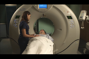
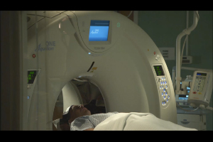
Each of the transplant recipients underwent preoperative CT with 3-D visualization. To build each life-size skull model, the CT images of the transplant recipient's head were segmented and processed using customized software, creating specialized data files that were input into a 3-D printer.
"In some patients, we need to modify the recipient's facial bones prior to transplantation," Dr. Imanzadeh said. "The 3-D printed model helps us to prepare the facial structures so when the actual transplantation occurs, the surgery goes more smoothly."
Although the entire transplant procedure lasts as long as 25 hours, the actual vascular connections from the donor face to the recipient typically takes approximately one hour, during which time the patient's blood flow must be stopped.
"If there are absent or missing bony structures needed for reconstruction, we can make modifications based on the 3-D printed model prior to the actual transplantation, instead of taking the time to do alterations during ischemia time," Dr. Rybicki said. "The 3-D model is important for making the transplant cosmetically appealing."
The researchers said they also used the models in the operating room to increase the surgeons' understanding of the anatomy of the recipient's face during the procedure.
"You can spin, rotate and scroll through as many CT images as you want but there's no substitute for having the real thing in your hand," Dr. Rybicki said. "The ability to work with the model gives you an unprecedented level of reassurance and confidence in the procedure."
Senior surgeons and radiologists involved in the five face transplantations agreed that the 3-D printed models provided superior pre-operative data and allowed complex anatomy and bony defects to be better appreciated, reducing total procedure time.
"Less time spent in the operating room is better for overall patient outcomes," Dr. Pomahac added.
Based on the results of this study, 3-D printing is now routinely used for surgical planning for face transplantation procedures at Brigham and Women's Hospital, and 3-D printed models may be implemented in other complex surgeries.
Co-authors on the study are Maximilian Kueckelhaus, M.D., Kanako K. Kumamaru, M.D., Ph.D., Nicole Wake, M.S., Dimitris Mitsouras, Ph.D., Elizabeth George, M.D., Gerald T. Grant, D.M.D., M.S., Peter C. Liacouras, Ph.D., and Edward J. Caterson, M.D., Ph.D.
Note: Copies of RSNA 2014 news releases and electronic images will be available online at RSNA.org/press14 beginning Monday, Dec. 1.
RSNA is an association of more than 54,000 radiologists, radiation oncologists, medical physicists and related scientists, promoting excellence in patient care and health care delivery through education, research and technologic innovation. The Society is based in Oak Brook, Ill. (RSNA.org)
Editor's note: The data in these releases may differ from those in the published abstract and those actually presented at the meeting, as researchers continue to update their data right up until the meeting. To ensure you are using the most up-to-date information, please call the RSNA Newsroom at 1-312-791-6610.
For patient-friendly information on CT of the head, visit RadiologyInfo.org.
Images (.JPG and .TIF format)
and Video clips (.mp4 format)

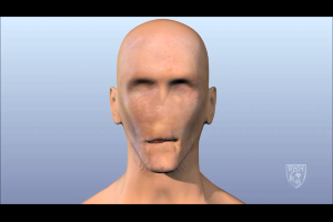
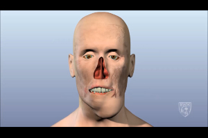
Video 1. Footage showing animation of face transplant process on a male patient.
Download.mp4
(Right-click and Save As)
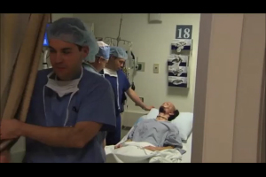
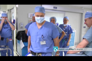
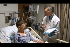
Video 2. Footage showing doctor and patient consultation prior to surgery.
Download.mp4
(Right-click and Save As)
Video 4. Footage showing doctor carrying cooler containing face organ
Download.mp4
(Right-click and Save As)
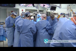
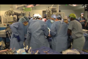
Video 5. Footage showing multiple doctors performing transplant.
Download.mp4
(Right-click and Save As)
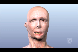
Video 6. Footage showing animation of face transplant process on a female patient.
Download.mp4
(Right-click and Save As)
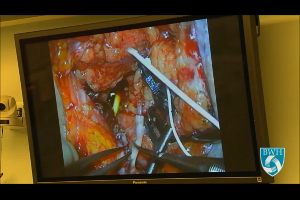
Video 7. Footage showing close-up of facial tissue, veins and blood cells while doctors perform surgery.
Download.mp4
(Right-click and Save As)
Video 8. Footage showing animation of face transplant process on another male patient.
Download.mp4
(Right-click and Save As)
Video 9. Footage showing doctor and patient consultation prior to surgery.
Download.mp4
(Right-click and Save As)
Video 12. Dr. Frank Rybicki explains the information that 3-D printing provides.
Download.mp4
(Right-click and Save As)
Video 13. Dr. Frank Rybicki explains why CT is critical to making the 3-D model.
Download.mp4
(Right-click and Save As)
Video 14. Dr. Frank Rybicki explains other medical and surgical uses for 3-D models.
Download.mp4
(Right-click and Save As)
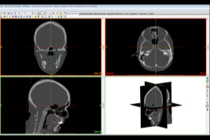
Video 15. CT Reconstruction for 3D Printing (Video provided by Materialise and Mimics Software)
Download.mp4
(Right-click and Save As)