RSNA Press Release
- Brain iron levels in patients with ADHD can be evaluated using an MRI technique called magnetic field correlation imaging.
- The results showed that ADHD patients who had never been on medication had lower brain iron levels compared to treated patients and the control group.
- The findings suggest that iron absorption into the brain may be abnormal in ADHD patients.
MRI Technique May Help Prevent ADHD Misdiagnosis
Released: June 17, 2014
| Media Contacts: | |
| RSNA Media Relations: | 1-630-590-7762 |
| Linda Brooks 1-630-590-7738 lbrooks@rsna.org |
Maureen Morley 1-630-590-7754 mmorley@rsna.org |
OAK BROOK, Ill. – Brain iron levels offer a potential biomarker in the diagnosis of attention deficit hyperactivity disorder (ADHD) and may help physicians and parents make better informed treatment decisions, according to new research published online in the journal Radiology.
ADHD is a common disorder in children and adolescents that can continue into adulthood. Symptoms include hyperactivity and difficulty staying focused, paying attention and controlling behavior. The American Psychiatric Association reports that ADHD affects 3 to 7 percent of school-age children.

Psychostimulant medications such as Ritalin are among the drugs commonly used to reduce ADHD symptoms. Psychostimulants affect levels of dopamine, a neurotransmitter in the brain associated with addiction.
"Much debate and concern has emerged regarding the continual rise of ADHD diagnosis in the U.S. given that two-thirds of those diagnosed receive psychostimulant medications," said Vitria Adisetiyo, Ph.D., postdoctoral research fellow at the Medical University of South Carolina in Charleston, S.C. "We wanted to see if we could identify brain iron as a potential noninvasive biomarker for medication-naïve ADHD to prevent misdiagnosis."
For the study, the research team measured brain iron levels in 22 children and adolescents with ADHD, 12 of whom had never been on medication for their condition (medication naïve), and 27 healthy control children and adolescents using a magnetic resonance imaging (MRI) technique called magnetic field correlation imaging. The technique was introduced in 2006 by study co-authors and faculty members Joseph A. Helpern, Ph.D., and Jens H. Jensen, Ph.D. No contrast agents were used, and blood iron levels in the body were measured using a blood draw.
The results showed that the 12 ADHD medication-naïve patients had significantly lower brain iron levels than the 10 ADHD patients who had been on psychostimulant medication and the 27 children and adolescents in the control group. In contrast, ADHD patients with a history of psychostimulant medication treatment had brain iron levels comparable to controls, suggesting that brain iron may increase to normal levels with psychostimulant treatment.
"Our research suggests that iron absorption into the brain may be abnormal in ADHD given that atypical brain iron levels are found even when blood iron levels in the body are normal," Dr. Adisetiyo said. "We found no differences in blood iron measures between controls, medication-naïve ADHD patients or pscyhostimulant-medicated ADHD patients."
Magnetic field correlation imaging's ability to noninvasively detect the low iron levels may help improve ADHD diagnosis and guide optimal treatment. Currently, ADHD diagnosis is based only on subjective clinical interviews and questionnaires. Having a biological biomarker may help inform clinical diagnosis, particularly in borderline cases, Dr. Adisetiyo noted.
If the results can be replicated in larger studies, magnetic field correlation might have a future role in determining which patients would benefit from psychostimulants—an important consideration because the drugs can become addictive if taken inappropriately and lead to abuse of other drugs like cocaine.
"We want the public to know that progress is being made in identifying potential noninvasive biological biomarkers of ADHD which may help to prevent misdiagnosis," Dr. Adisetiyo said. "We are currently testing our findings in a larger cohort to confirm that measuring brain iron levels in ADHD is indeed a reliable and clinically feasible biomarker."
# # #
"Multimodal MR Imaging of Brain Iron in Attention Deficit Hyperactivity Disorder: A Non-invasive Biomarker that Responds to Psychostimulant Treatment?" Collaborating with Drs. Adisetiyo, Helpern and Jensen were Ali Tabesh, Ph.D., Rachael L. Deardorff, M.S., Els Fieremans, Ph.D., Adriana Di Martino, M.D., Kevin M. Gray, M.D., and Francisco X. Castellanos, M.D. Study abstract
Radiology is edited by Herbert Y. Kressel, M.D., Harvard Medical School, Boston, Mass., and owned and published by the Radiological Society of North America, Inc. (Radiology.RSNA.org/)
RSNA is an association of more than 53,000 radiologists, radiation oncologists, medical physicists and related scientists, promoting excellence in patient care and health care delivery through education, research and technologic innovation. The Society is based in Oak Brook, Ill. (RSNA.org)
For patient-friendly information on MRI of the brain, visit RadiologyInfo.org.
Images (.JPG and .TIF format) and Video clips (.mp4 format)
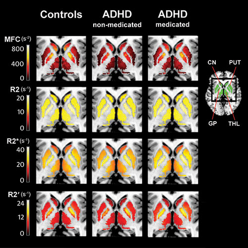 Figure 1. Subgroup averages of parametric maps of MRI brain iron indices: magnetic field correlation (MFC: top row) and relaxation rates R2 (second row), R2*(third row), and R2' (bottom row). Parametric maps, masked for regions of interests (green): caudate nucleus (CN), putamen (PUT), globus pallidus (GP) and thalamus (THL), are shown for controls (n = 27), medication-naïve ADHD patients (ADHD-non-medicated; n = 12) and ADHD patients with a history of psychostimulant treatment (ADHD-medicated; n = 10). The ADHD-non-medicated subgroup displayed significantly reduced striatal (CN, PUT) and thalamic MFC compared to both controls and the ADHD-medicated subgroup. No significant differences were detected between the latter two groups. High-res (TIF) version (Right-click and Save As) |
 Video 1. Dr. Vitria Adisetiyo explains why her research team conducted this research. Download.mp4 (Right-click and Save As) |
 Video 2. Dr. Vitria Adisetiyo discusses the most exciting finding of her research. Download.mp4 (Right-click and Save As) |
 Video 3. Dr. Vitria Adisetiyo explains magnetic field correlation. Download.mp4 (Right-click and Save As) |
 Video 4. Dr. Vitria Adisetiyo discusses the advantages of magnetic field correlation. Download.mp4 (Right-click and Save As) |
|
 Video 5. Dr. Vitria Adisetiyo discusses the risks of prescribing Ritalin to children with normal brain iron levels. Download.mp4 (Right-click and Save As) |
 Video 6. Dr. Vitria Adisetiyo explains how their research may help diagnose children with ADHD. Download.mp4 (Right-click and Save As) |
 Video 7. Dr. Vitria Adisetiyo discusses the next step in their research. Download.mp4 (Right-click and Save As) |
 PDF
PDF