RSNA Press Release
- Researchers using MRI have found that Alzheimer’s disease causes a different pattern of brain volume loss in men, compared to women.
- Women with AD initially show greater brain atrophy, but the disease progresses more quickly in men.
- Approximately 5.4 million Americans have AD.
Researchers Discover Gender-based Differences in Alzheimer’s Disease
Released: November 26, 2012
| Media Contacts: | RSNA Newsroom | 1-312-949-3233 |
| Before 11/24/12 or after 11/29/12: | RSNA Media Relations: | 1-630-590-7762 |
| |
Linda Brooks 1-630-590-7738 lbrooks@rsna.org |
Maureen Morley 1-630-590-7754 mmorley@rsna.org |
CHICAGO—All patients with Alzheimer's disease (AD) lose brain cells, which leads to a shrinking, or atrophy, of the brain. But the pattern of gray matter loss is significantly different in men and women, according to a study presented today at the annual meeting of the Radiological Society of North America (RSNA).

"We found that the extent and distribution of regional gray matter volume loss in the brain was strongly influenced by gender," said lead researcher Maria Vittoria Spampinato, M.D., associate professor of radiology at the Medical University of South Carolina in Charleston.
According to the Alzheimer's Association, 5.4 million Americans have AD, the sixth-leading cause of death in the U.S. Currently, there is no cure for AD, which lends urgency to research efforts designed to better understand, diagnose and treat this devastating illness.
"There is a strong interest in using magnetic resonance imaging (MRI) to assess brain atrophy with the purpose of monitoring dementia progression noninvasively and to aid in understanding which factors can influence brain atrophy progression and distribution in the Alzheimer's brain," Dr. Spampinato said.
In the study, Dr. Spampinato and colleagues analyzed data on 109 patients, including 60 men and 49 women (mean age 77), who participated in the Alzheimer's Disease Neuroimaging Initiative (ADNI), a major study that followed hundreds of cognitively healthy individuals and individuals with mild cognitive impairment (MCI) and AD over a period of five years.
During the five-year period, each of the 109 patients progressed from amnestic MCI (in which the patient suffers memory loss but maintains cognitive function) to AD. Using MR images of the patients' brains taken when they were diagnosed with AD and 12 months before and after the diagnosis, the researchers created brain maps that illustrated gray matter changes.
The brain maps revealed that compared to male patients, the women had greater atrophy in gray matter 12 months prior to their AD diagnosis and at the time of their diagnosis. The brain maps also showed that the men and women in the study lost gray matter volume in different areas of the brain as their disease progressed from MCI to AD.
"The female patients in our study initially had more gray matter atrophy than the male patients but over time, the men caught up," Dr. Spampinato said. "In the men, the disease developed more aggressively in a shorter period of time."
Dr. Spampinato said the gender differences in atrophy patterns have important implications for the development of therapies for MCI and AD.
"These differences should be taken into consideration when testing new drugs in clinical trials," she said. "Knowing the difference between the male and female patterns of atrophy will help researchers better decipher a patient's response to drug therapy."
Coauthors are Zoran Rumboldt, M.D., Markus Weininger, M.D., Hrvoje Vavro, M.D., Karen Patrick, M.D., and Ryan O'Neal Parker, Ph.D.
# # #
Note: Copies of RSNA 2012 news releases and electronic images will be available online at RSNA.org/press12 beginning Monday, Nov. 26.
RSNA is an association of more than 50,000 radiologists, radiation oncologists, medical physicists and related scientists promoting excellence in patient care and health care delivery through education, research and technologic innovation. The Society is based in Oak Brook, Ill. (RSNA.org)
Editor's note: The data in these releases may differ from those in the published abstract and those actually presented at the meeting, as researchers continue to update their data right up until the meeting. To ensure you are using the most up-to-date information, please call the RSNA Newsroom at 1-312-949-3233.
For patient-friendly information on MRI of the brain, visit RadiologyInfo.org.
| Abstract: |
Video clips
- (.mp4 format)
- Video clip (17,075 Kbyte)
- Video clip (19,485 Kbyte)
- Video clip (34,627 Kbyte)
- Video clip (659 Kbyte)
Footage showing the view from the control room during a magnetic resonance imaging (MRI) scan. - Video clip (172 Kbyte)
Footage showing a patient entering a magnetic resonance imaging (MRI) scanner. - Video clip (616 Kbyte)
Footage showing a radiologic technologist preparing a patient for a magnetic resonance imaging (MRI) scan and the view from the control room. - Video clip (362 Kbyte)
Footage showing the view from the control room during a magnetic resonance imaging (MRI) scan and brain MR images.
Images (.JPG format)
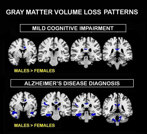 Figure 1. Shown in blue are areas of relatively greater brain volume in males compared to females 12 months before Alzheimer’s disease diagnosis (Mild Cognitive Impairment) and at the time of Alzheimer’s disease diagnosis (Alzheimer’s disease diagnosis). No areas of greater brain volume were found in females than males. High-res (TIF) version (Right-click and Save As) |
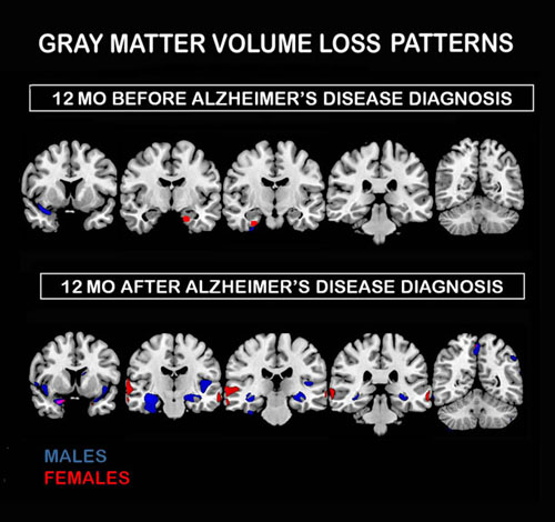 Figure 2. Shown are areas of brain volume loss found in males (blue) and in females (red) during the 12 months before and during the 12 months after Alzheimer’s disease diagnosis (common areas of volume loss in purple). High-res (TIF) version (Right-click and Save As) |
 Figure 3. Photograph of a Siemens Magnetom Avanto 1.5T MRI Scanner. High-res (TIF) version (Right-click and Save As) |
 PDF
PDF