RSNA Press Release
- Imaging is showing new and unusual injuries to the arms, wrists and hands of adolescent gymnasts.
- The study found that some of the gymnasts experience "early death" of a portion of their knuckle bones.
- Adolescent gymnasts with overuse injuries to the wrists, hands and knuckles will likely develop early osteoarthritis.
MRI Shows New Types of Injuries in Young Gymnasts
Released: December 1, 2008
| Media Contacts: | RSNA Newsroom | 1-312-949-3233 |
| Before 11/29/08 or after 12/04/08: | RSNA Media Relations: | (630) 590-7762 |
| |
Maureen Morley 1-630-590-7754 mmorley@rsna.org |
Linda Brooks 1-630-590-7738 lbrooks@rsna.org |
CHICAGO — Adolescent gymnasts are developing a wide variety of arm, wrist and hand injuries that are beyond the scope of previously described gymnastic-related trauma, according to a study presented today at the annual meeting of the Radiological Society of North America (RSNA).
"The broad constellation of recent injuries is unusual and might point to something new going on in gymnastics training that is affecting young athletes in different ways," said the study's lead author, Jerry Dwek, M.D., an assistant clinical professor of radiology at the University of California, San Diego and a partner of San Diego Imaging at Rady Children’s Hospital and Health Center.
Previous studies have reported on numerous injuries to the growing portion of adolescent gymnasts' bones. However, this study uncovered some injuries to the bones in the wrists and knuckles that have not been previously described. In addition, the researchers noted that these gymnasts had necrosis, or "early death," of the bones of their knuckles.
"These young athletes are putting an enormous amount of stress on their joints and possibly ruining them for the future," Dr. Dwek said.
The radius is the bone in the forearm that takes the most stress during gymnastics. Due to damage to the radial growth plates, the bone does not grow in proportion to the rest of the skeleton and may be deformed. Consequently, it is not unusual for gymnasts to have a longer ulna than radius. Some former gymnasts must undergo surgery to shorten the ulna and regain the proper fit of the wrist bones into the forearm.
Dr. Dwek and coauthor Christine Chung, M.D., used MRI to study overuse injuries seen in the skeletally immature wrists and hands of gymnasts. The researchers studied wrist and hand images of 125 patients, age 12 to 16, including 12 gymnasts with chronic wrist or hand pain.
"We were surprised to be looking at injuries every step down the hand all the way from the radius to the small bones in the wrist and on to the ends of the finger bones at the knuckles," Dr. Dwek said. "These types of injuries are likely to develop into early osteoarthritis."
Dr. Dwek suggested that additional study is needed to understand how gymnastic stresses are causing these injuries.
"It is possible that by changing the way that practice routines are performed, we might be able to limit the stress on the joints and on delicate growing bones," he said.
# # #
RSNA is an association of more than 42,000 radiologists, radiation oncologists, medical physicists and related scientists committed to excellence in patient care through education and research. The Society is based in Oak Brook, Ill. (RSNA.org)
Editor's note: The data in these releases may differ from those in the printed abstract and those actually presented at the meeting, as researchers continue to update their data right up until the meeting. To ensure you are using the most up-to-date information, please call the RSNA Newsroom at 1-312-949-3233.
For patient-friendly information on MRI, visit RadiologyInfo.org.
| Abstract: |
Images (.JPG format)
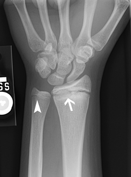 Figure 1. An x-ray image illustrating an irregular and widened growth plate of the hand (arrow). The irregularity is caused by repetitive trauma and can result in shortening and deformity. High-res (TIF) version (Right-click and Save As) |
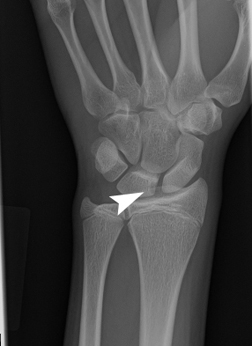 Figure 2. An x-ray image illustrating a lunate bone with a defect in its form, indicating a dead bone. This injury has not been reported previously in gymnasts. High-res (TIF) version (Right-click and Save As) |
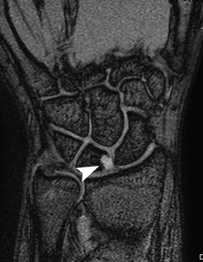 Figure 3. An MRI image illustrating focal lunate necrosis (death of a small portion of the lunate bone). High-res (TIF) version (Right-click and Save As) |
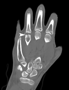 Figure 4. A CT scan illustrating flattening and irregularity at the end of the fourth metacarpal, a bone that forms knuckles in the hand. High-res (TIF) version (Right-click and Save As) |
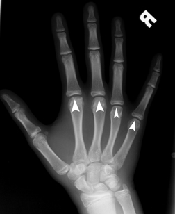 Figure 5. An MRI image illustrating flattening of the heads of the metacarpals (bones that form the knuckles of the hands). This injury has not been described previously in gymnasts. High-res (TIF) version (Right-click and Save As) |
 Figure 6. 1.5 Tesla MRI machine. High-res (TIF) version (Right-click and Save As) |
|
|
 PDF
PDF