RSNA Press Release
- Abnormalities in language processing in children with autism can be identified using magnetoencephalography (MEG).
- The brain's auditory processing system is delayed a fraction of a second in children with autism.
- Autism affects one in every 150 American children, mostly boys.
Brain Waves Show Sound Processing Abnormalities in Autistic Children
Released: December 1, 2008
| Media Contacts: | RSNA Newsroom | 1-312-949-3233 |
| Before 11/29/08 or after 12/04/08: | RSNA Media Relations: | (630) 590-7762 |
| |
Maureen Morley 1-630-590-7754 mmorley@rsna.org |
Linda Brooks 1-630-590-7738 lbrooks@rsna.org |
CHICAGO — Abnormalities in auditory and language processing may be evaluated in children with autism spectrum disorder by using magnetoencephalography (MEG), according to a study presented today at the annual meeting of the Radiological Society of North America (RSNA).
"Using MEG, we can record the tiny magnetic fields associated with electrical brain activity," said Timothy Roberts, Ph.D., vice chair of research in the Department of Radiology at Children’s Hospital of Philadelphia. "Recorded brain waves change with every sensation, thought and activity. It's like watching a movie of the brain in real time."
Typically used for epilepsy evaluation, MEG can also be used to identify timing abnormalities in the brains of patients with autism.
"We found that signatures of autism are revealed in the timing of brain activity," Dr. Roberts said. "We see a fraction of a second delay in autistic patients."
Autism is a complex developmental disability that affects approximately one in every 150 American children, mostly boys, according to the Autism Society of America. Autism inhibits the brain functions that govern the development of social and communication skills.
For a MEG exam, a helmet that houses magnetic detectors and looks similar to an old-fashioned hair dryer is lowered over the patient's head while the patient remains in a seated position. The helmet analyzes electrical currents from the brain.
For the study, 64 patients, age six to 15, with a diagnosis of autism spectrum disorder were evaluated with MEG. Audio stimulation was introduced to the children in the form of beeps, tones in pairs, vowels or sentences. Sounds were presented at different frequencies and tone pairs in rapid succession, including unusual streams of incongruous tones and vowels. The results were analyzed and compared with the results from a control group of age-matched non-autistic children.
The findings showed that in the children with autism there was a fraction of a second delay in the brain's response while processing the rapid succession sounds and the unusual streams, giving researchers an insight into the dysfunction of the auditory processing system in autistic children.
"This delay in processing certain types and streams of sound may underpin the subsequent language processing and communication impairment seen in autistic children," Dr. Roberts said.
Dr. Roberts predicts that the signatures of autism found in brain activity will become biomarkers to improve classification of the disorder and aid in treatment and therapy planning.
"We hope that in the future these signatures will also be revealed in the infant brain to help diagnose autism and allow earlier intervention," he said.
Co-authors are J. Christopher Edgar, Ph.D., Deborah M. Zarnow, M.D., and Susan E. Levy, M.D.
# # #
Disclosure: This study was funded by the National Institutes of Health and by the Nancy Lurie Marks Family Foundation.
RSNA is an association of more than 42,000 radiologists, radiation oncologists, medical physicists and related scientists committed to excellence in patient care through education and research. The Society is based in Oak Brook, Ill. (RSNA.org)
Editor's note: The data in these releases may differ from those in the printed abstract and those actually presented at the meeting, as researchers continue to update their data right up until the meeting. To ensure you are using the most up-to-date information, please call the RSNA Newsroom at 1-312-949-3233.
For patient-friendly information on imaging technologies and procedures, visit RadiologyInfo.org.
| Abstract: |
Images (.JPG format)
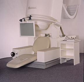 Figure 1. Depiction of a biomagnetometer. High-res (TIF) version (Right-click and Save As) |
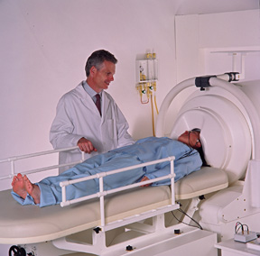 Figure 2. Depiction of a biomagnetometer with a patient in the horizontal position. High-res (TIF) version (Right-click and Save As) |
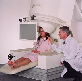 Figure 3. Depiction of a biomagnetometer with a patient in the upright position. High-res (TIF) version (Right-click and Save As) |
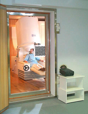 Figure 4. Depiction of a biomagnetometer. High-res (TIF) version (Right-click and Save As) |
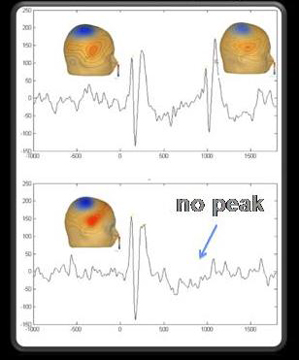 Figure 5. This MEG image depicts the response of an autistic child with language impairment. High-res (TIF) version (Right-click and Save As) |
 Figure 6. When children with autism hear tones in rapid succession (lower left graph), their response to the second tone is greatly reduced and delayed compared to responses by typically developing children (lower right graph). Upper graphs show responses to two tones separated at a longer interval. Blue and red colors on the skull diagrams indicate signal strengths. High-res (TIF) version (Right-click and Save As) |
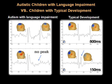 Figure 7. When children with autism hear tones in rapid succession (lower left graph), their response to the second tone is greatly reduced and delayed compared to responses by typically developing children (lower right graph). Upper graphs show responses to two tones separated at a longer interval. Blue and red colors on the skull diagrams indicate signal strengths. High-res (TIF) version (Right-click and Save As) |
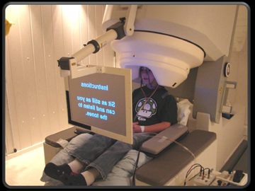 Figure 8. A patient reads instructions on a screen while seated with his head surrounded by the MEG's magnetic detectors. High-res (TIF) version (Right-click and Save As) |
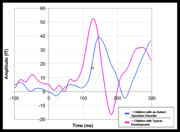 Figure 9. Compared to a typically developing child (magenta curve), an autistic child's response (blue) to a sound is weaker and delayed in time by a fraction of second. High-res (TIF) version (Right-click and Save As) |
 Figure 10. This MEG image depicts the response of a typically developing child. High-res (TIF) version (Right-click and Save As) |
 PDF
PDF