RSNA Press Release
- Memorizing a long poem or newspaper article can improve verbal and episodic recall in older adults.
- Changes in brain chemistry and memory are not evident immediately after rote learning but appear after a lengthy period of rest.
- The brain should be exercised as a defense against dementia, cognitive lapses and memory failure.
Rote Learning Improves Memory in Seniors
Released: November 27, 2006
| Media Contacts: | ||
| RSNA Media Relations: | (630) 590-7762 | |
| Maureen Morley (630) 590-7754 mmorley@rsna.org |
||
CHICAGO — A new study offers older adults a simple way to combat memory loss: memorization. Researchers found that seniors who engaged in an intensive period of rote learning followed by an equally long rest period exhibited improved memory and verbal recall. The study was presented today at the annual meeting of the Radiological Society of North America (RSNA).
"We didn't see an immediate improvement following the intensive memorization period," said Jonathan McNulty, B.Sc., H.Dip., of Diagnostic Imaging at the School of Medicine and Medical Science, University College Dublin in Ireland. "However, after a six-week rest, the volunteers manifested both metabolic changes in the brain and improved memory performance."
As people age, they often begin to experience forgetfulness and may have difficulty learning new material. Approximately 40 percent of people over age 60 have some kind of memory difficulty. Mild, age-related memory loss is caused by the loss of brain cells over time, along with changes in brain chemistry.
The researchers studied how repeated cognitive exercise impacts memory and recall, as well as the health of brain cells involved in memory.
The study involved 24 healthy older adults between the ages of 55 and 70. The volunteers engaged in six weeks of intensive rote learning, memorizing a newspaper article or poem of 500 words, followed by six weeks of rest.
An extensive battery of learning and memory tests was administered before and after the six-week learning period. Magnetic resonance spectroscopy (MRS), a special type of magnetic resonance imaging, was performed on half of the volunteers before and after the intensive learning session, and again six weeks later. MRS was used to measure changes in N-acetylaspartate, creatine and choline, three metabolites in the brain that are related to memory performance and neural cell health.
At the end of the six-week learning session, no changes in the brain metabolism or memory performance were observed. But following the rest period, all of the volunteers experienced improvements in their verbal and episodic memory—they were better able to remember and repeat a short story and a list of words and to recall events that occurred earlier in the day or week. These behavioral changes correlated with metabolic changes identified by MRS in the left posterior hippocampus, a memory-related brain structure.
"Unlike other studies on memory involving specific training regimes, memorizing is an everyday activity that anyone can undertake," said co-author Richard Roche, Ph.D., of the Department of Psychology at National University of Ireland in Maynooth. "The brain is like a muscle that should be exercised through the retirement years as a defense against dementia, cognitive lapses and memory failure."
Co-authors are Paul Brennan, M.D., Colin P. Doherty, M.D., D. McMackin, M.D., S. Sukumaran, M.D., I.H. Robertson, Ph.D., M.A. Mangaoang, Ph.D., S.M. O'Mara, D.Phil., Sinead L. Mullally, Ph.D., J. Hayden, B.A., J. Prendergast, B.Sc., and M. Fitzsimons, Ph.D.
# # #
RSNA is an association of more than 40,000 radiologists, radiation oncologists, medical physicists and related scientists committed to promoting excellence in radiology through education and by fostering research, with the ultimate goal of improving patient care. The Society is based in Oak Brook, Ill.
Editor's note: The data in these releases may differ from those in the printed abstract and those actually presented at the meeting, as researchers continue to update their data right up until the meeting. To ensure you are using the most up-to-date information, please call the RSNA Newsroom at (312) 949-3233.
| Abstract: |
Images (.JPG format)
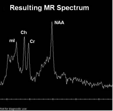 Figure 1. This image shows what a normal MR spectrum might look like. It is a graph of signal intensity versus frequency (parts per million) of the various metabolites. |
High-res (TIF) version (Right-click and Save As) |
|
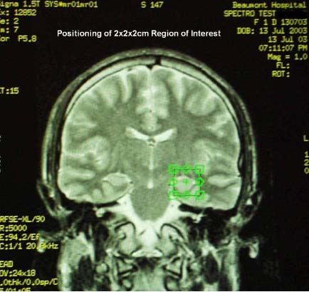 Figure 2. This image shows a coronal slice through the brain and the positioning of one of the seven regions of interest used to produce spectra. Each region or voxel is 2cm3. |
High-res (TIF) version (Right-click and Save As) |
|
 PDF
PDF