RSNA Press Release
- CT images of King Tut's mummy show that a thigh wound may have precipitated his death.
- Researchers may have disproved previous claims that Tut was killed by a blow to the head.
- The investigation did not escape Tut's "curse," suffering mishaps including power losses and the illness of a team member.
Radiologists Attempt to Solve Mystery of Tut's Demise
Released: November 27, 2006
CHICAGO — Egyptian radiologists who performed the first-ever computed tomography (CT) evaluation of King Tutankhamun's mummy believe they have solved the mystery of how the ancient pharaoh died. The CT images and results of their study were presented today at the annual meeting of the Radiological Society of North America (RSNA).
Ashraf Selim, M.D., radiologist at Kasr Eleini Teaching Hospital, Cairo University in Egypt, was part of an international team of scientists that studied the 3,300-year-old mummy of King Tut in Egypt. Using a mobile multi-detector CT scanner, the researchers performed a full-body scan on the king's remains, obtaining approximately 1,900 digital cross-sectional images.
"We found the mummy was in a critical stage of preservation," said Dr. Selim. "The body was cut into several parts with some missing pieces."
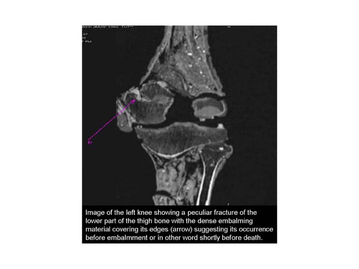
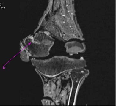
With the help of the CT images, researchers estimated King Tut's age at death to be between 18 and 20 years. His height was 180 centimeters or approximately 5 feet 11 inches. The researchers discovered a possible premortem fracture to the femoral (thigh) bone. While they cannot assess how the injury occurred, the findings suggest that the injury may have been an open wound that became infected and ultimately fatal.
Since King Tut was first examined by x-ray in 1968, revealing what appeared to be a bone fragment in his skull, it has been widely speculated that a blow to the head killed the boy king. However, Dr. Selim and colleagues found several pieces of evidence to the contrary. In the cranial cavity, they found loose bone fragments that were not covered with the intracranial solidified embalming material. These bone fragments matched exactly a defect within the first vertebra in the neck. They found no evidence of skull fractures.
A mishap during the mummification process, or even damage incurred during that first x-ray examination may explain the misplaced—and misleading—bone fragments. Dr. Selim suggests the damage may have been caused by the expedition led by Howard Carter that first discovered Tut's tomb in 1922.
"We believe that this broken piece from the first vertebra of the king's spine may have been fractured and dislodged when Carter, Derry, Hamdy and their team tried to remove and free the gold mask, which was tightly glued and quite adherent to the body, by using some metal instruments that broke the thin, fragile piece of bone that lies immediately underneath the bone defect in the skull base through which the spinal cord emerges," Dr. Selim said.
Dr. Selim's team did not escape the so-called curse that is said to plague anyone who disrupts the remains of the boy king.
"While performing the CT scan of King Tut, we had several strange occurrences," he said. "The electricity suddenly went out, the CT scanner could not be started and a team member became ill. If we weren't scientists, we might have become believers in the Curse of the Pharaohs."
The CT examination of King Tut is part of a five-year initiative called the Egyptian Mummy Project to image and preserve Egypt's mummies and to solve various mysteries about the diseases and lifestyles of ancient Egyptians.
King Tutankhamun, who ascended to the throne when he was just eight years old, was mummified and buried with other ancient royals. His tomb, filled with 5,000 artifacts, was discovered near Luxor, Egypt in 1922. Artifacts from the tombs of King Tut and other royals buried in the Valley of the Kings are part of "Tutankhamun and the Golden Age of the Pharaohs," an exhibition currently at Chicago's Field Museum.
Co-authors of the study are Mervat Shafik, M.D., Essam Eisheikh, M.D., Sherif Abdel Fattah, M.D., Hany Amer, M.D., Zahi Hawass, D.S.C., A. Gamal Eldin, M.D., F. Gaballah, M.D., F. Ruhli, M.D. Ph.D., E. Egarter, M.D., and P. Gostner, M.D.
# # #
RSNA is an association of more than 40,000 radiologists, radiation oncologists, medical physicists and related scientists committed to promoting excellence in radiology through education and by fostering research, with the ultimate goal of improving patient care. The Society is based in Oak Brook, Ill.
Editor's note: The data in these releases may differ from those in the printed abstract and those actually presented at the meeting, as researchers continue to update their data right up until the meeting. To ensure you are using the most up-to-date information, please call the RSNA Newsroom at (312) 949-3233.
| Abstract: |
Video clips
- Video clip 1: CT series of King Tut's head (sagittal and coronal)
- Flash format (short download) (34 Kbyte)
- AVI format (long download) (3,147 Kbyte)
Images (.JPG format)
 (with caption) |
 (without caption) |
High-res (TIF) version (Right-click and Save As) |
|
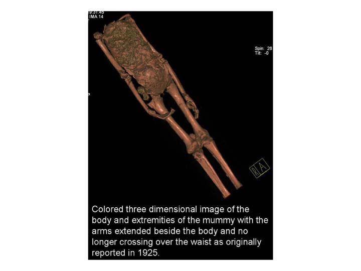
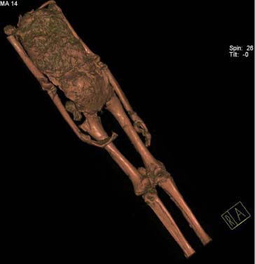
| Figure 1. Colored 3-D CT image of the body and extremities of the mummy with the arms extended beside the body, no longer crossing over the waist as originally reported in 1925. | |||
 (with caption) |
 (without caption) |
High-res (TIF) version (Right-click and Save As) |
|
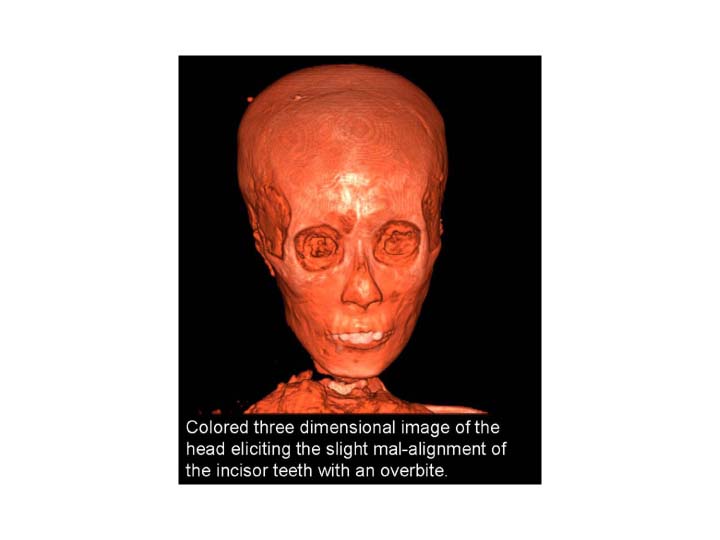
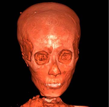
| Figure 2. Colored 3-D CT image of the head eliciting the slight mal-alignment of the incisor teeth with an overbite. | |||
 (with caption) |
 (without caption) |
High-res (TIF) version (Right-click and Save As) |
|
| Figure 3. CT image of the left knee showing a peculiar fracture of the lower part of the thigh bone with the dense embalming material covering its edges (arrow) suggesting its occurrence before embalmment, or in other words, shortly before death. | |||
 (with caption) |
(without caption) |
High-res (TIF) version (Right-click and Save As) |
|
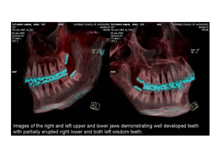
| Figure 4. Images of the right and left upper and lower jaws demonstrating well developed teeth with partially erupted right lower and both left wisdom teeth. | |||
 (with caption) |
 (without caption) |
High-res (TIF) version (Right-click and Save As) |
|
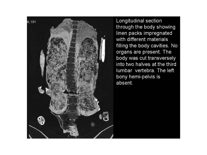
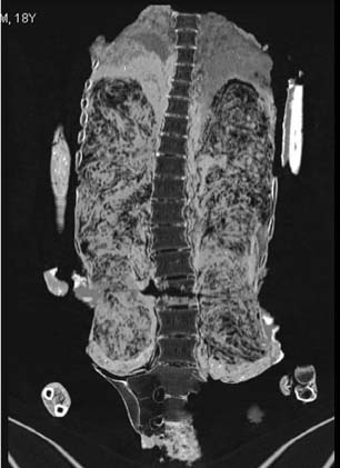
| Figure 5. Longitudinal section through the body showing linen packs impregnated with different materials filling the body cavities. No organs are present. The body was cut transversely into two halves at the third lumbar vertebra. The left bony hemi-pelvis is absent. | |||
 PDF
PDF