RSNA Press Release
- Researchers using diffusion tensor imaging (DTI) have found similar abnormalities in the brains of adolescents who are daily marijuana users and adolescents with schizophrenia.
- The abnormalities were found in a part of the brain still developing during adolescence that is associated with the higher aspects of language and auditory functions.
- The findings also suggest that heavy use of marijuana may lead to earlier onset of schizophrenia in adolescents who are genetically predisposed to the disorder.
Imaging Shows Similarities in Brains of Marijuana Smokers, Schizophrenics
Released: November 30, 2005
| Media Contacts: | |
| RSNA Media Relations: | (630) 590-7762 |
| Maureen Morley (630) 590-7754 mmorley@rsna.org |
Heather Babiar (630) 590-7738 hbabiar@rsna.org |
CHICAGO – Heavy use of marijuana may put adolescents who are genetically predisposed to schizophrenia at greater risk of developing the brain disorder, according to research presented today at the annual meeting of the Radiological Society of North America (RSNA).
Using a sophisticated brain imaging technique called diffusion tensor imaging (DTI), researchers at Zucker Hillside Hospital in Glen Oaks, New York, studied the brains of groups of adolescents: healthy, non-drug users; heavy marijuana smokers (daily use for at least one year); and schizophrenic patients. Unlike magnetic resonance imaging (MRI), which provides a static picture of brain structures, DTI detects and measures the motion of water molecules in the brain, which can reveal microscopic abnormalities.
Manzar Ashtari, Ph.D., Sanjiv Kumra, M.D., and colleagues used DTI to examine the arcuate fasciculus, a bundle of fibers connecting the Broca’s area in the left frontal lobe and the Wernicke's area in the left temporal lobe of the brain. The investigators found that repeated exposure to marijuana was related to abnormalities in the development of this fiber pathway, which is associated with the higher aspects of language and auditory functions.
"Because this language/auditory pathway continues to develop during adolescence, it is most susceptible to the neurotoxins introduced into the body through marijuana use," explained Dr. Ashtari, associate professor of radiology and psychiatry at New York's Albert Einstein College of Medicine.
In the study, DTI was performed on 12 healthy, early adolescent males compared with 12 late adolescent males to show normal human brain development; 11 schizophrenic patients compared with 17 matched controls; 15 schizophrenic patients who smoke marijuana compared with 17 matched controls; and 15 marijuana smokers compared with 15 matched non-drug users. The scans revealed no abnormal developmental changes in the language pathway in the healthy adolescents, but showed abnormalities in both the marijuana users and schizophrenic patients.
"These findings suggest that in addition to interfering with normal brain development, heavy marijuana use in adolescents may also lead to an earlier onset of schizophrenia in individuals who are genetically predisposed to the disorder," said co-principal-investigator Sanjiv Kumra, M.D., assistant professor of psychiatry at Albert Einstein College of Medicine.
According to the National Institute on Drug Abuse, approximately 3.1 million Americans age 12 and older use marijuana on a daily or almost daily basis. In 2004, 5.6 percent of 12 th graders reported daily use of marijuana.
Schizophrenia is a chronic, severe and disabling brain disorder that affects about one percent of the entire population. Although the causes of the disease have not been determined, it is believed to result from a combination of environmental and genetic factors.
Drs. Ashtari and Kumra said longitudinal studies are needed to determine whether these changes in the brain are permanent or change over time. It is also important to mention that at this time, DTI and MRI are not diagnostic means for schizophrenia patients or marijuana smokers.
Co-authors are Jinghui Wu, B.S., Kelly Cervellione, M.A., John Kane, M.D., Philip Szeszko, Ph.D., and Babak Ardekani, Ph.D.
# # #
Note: Copies of RSNA 2005 news releases and electronic images will be available online at RSNA.org/press05 beginning Monday, Nov. 28.
RSNA is an association of more than 38,000 radiologists, radiation oncologists, medical physicists and related scientists committed to promoting excellence in radiology through education and by fostering research, with the ultimate goal of improving patient care. The Society is based in Oak Brook, Ill.
Editor's note: The data in these releases may differ from those in the printed abstract and those actually presented at the meeting, as researchers continue to update their data right up until the meeting. To ensure you are using the most up-to-date information, please call the RSNA Newsroom at (312) 949-3233.
| Abstract: |
Images (.JPG format)
Figure 1. Water motion along the tail of brain cells (axons). |
 Figure 2. T1 Weighted MRI of brain. |
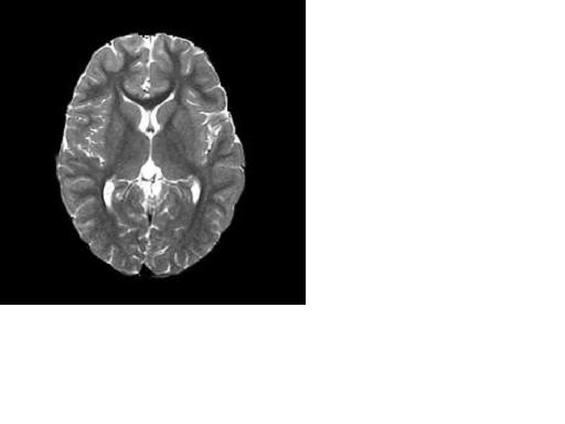 Figure 3. T2 Weighted MRI of brain. |
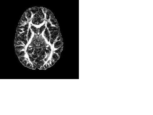 Figure 4. DTI of brain (FA Map). Hi-res (TIF) version (Right-click and Save As) |
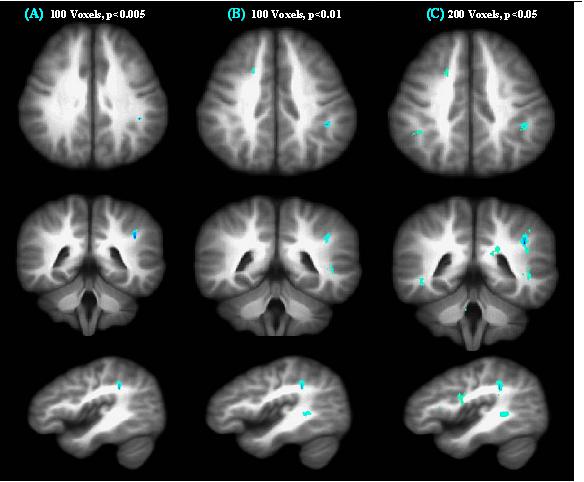 Figure 5. Normal brain development, blue clusters are developed during late adolescent years. |
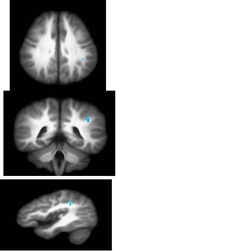 Figure 6. images from Normals; blue clusters are brain under construction. |
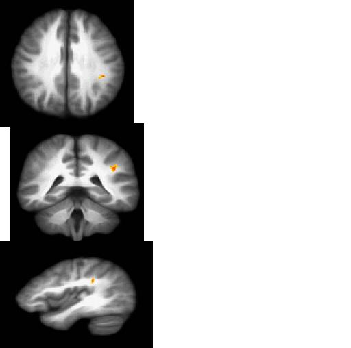 Figure 7. Images from Marijuana users; orange clusters are structurally altered brain areas. Hi-res (TIF) version (Right-click and Save As) |
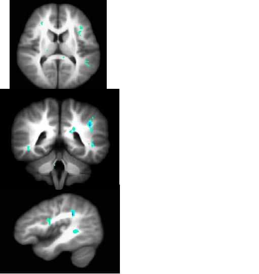 Figure 8. images from Normals study; blue clusters are brain under construction. Hi-res (TIF) version (Right-click and Save As) |
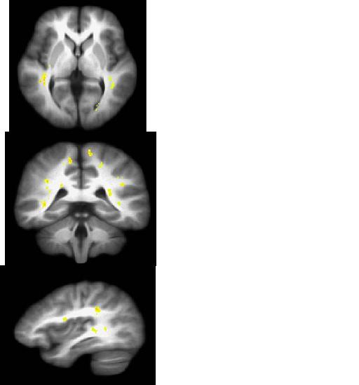 Figure 9. Images from Schizophrenia study; yellow clusters are brain damaged areas. Hi-res (TIF) version (Right-click and Save As) |
 PDF
PDF