RSNA Press Release
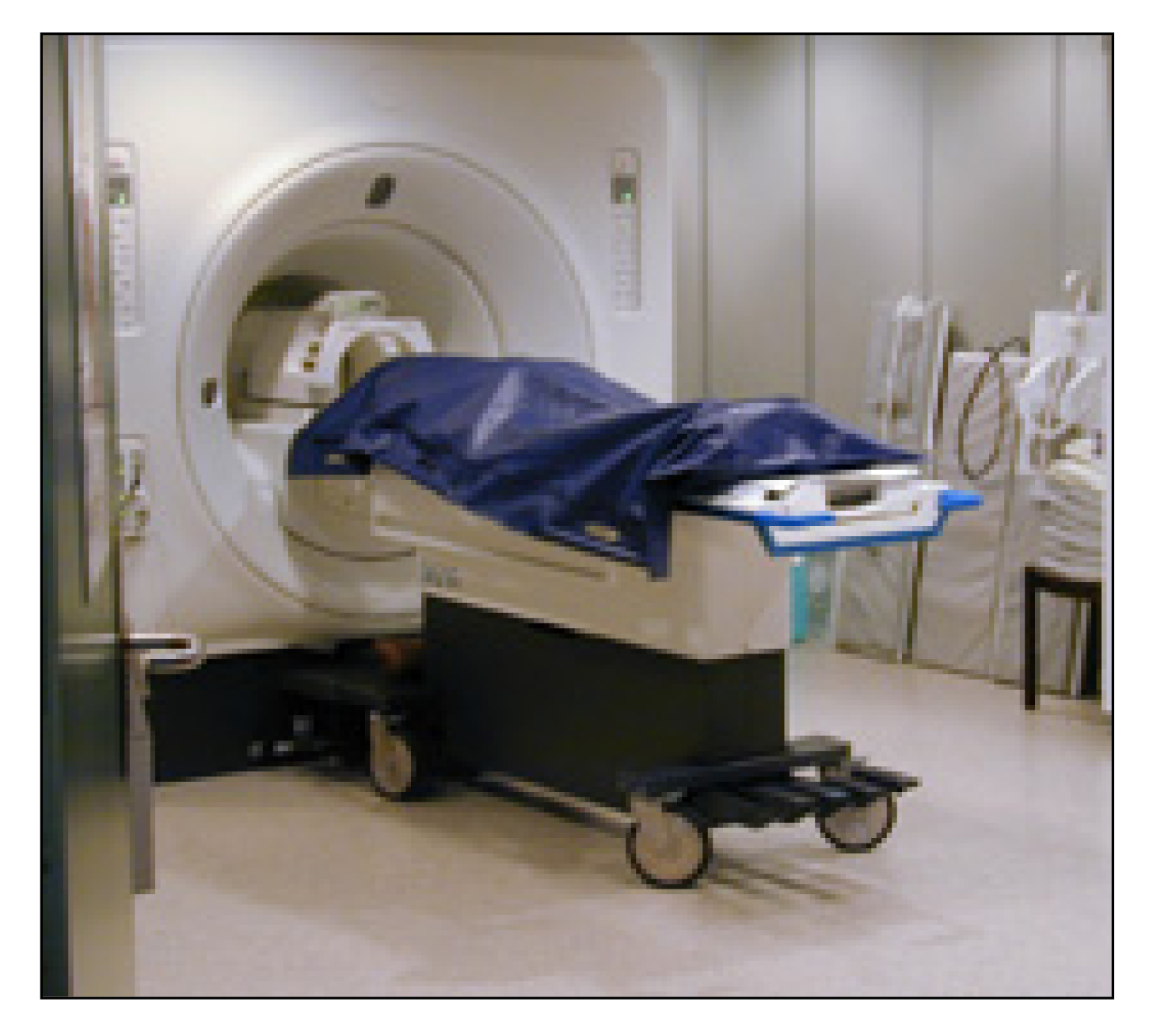
- Combining computed tomography (CT) and magnetic resonance (MR) imaging, Swiss researchers are conducting bloodless, non-invasive virtual autopsies.
- Virtual autopsy does not destroy evidence and can be used in situations where religious beliefs or families reject the traditional autopsy.
- 3-D virtual autopsy images eliminate the need for juries to view graphic photographs.
Forensic Radiology Makes Virtual Autopsy a Reality
Released: December 3, 2003
|
Media Contacts: |
Heather Babiar or Maureen Morley | (630) 590-7762 |
| Heather Babiar (630) 590-7738 hbabiar@rsna.org |
Maureen Morley (630) 590-7754 mmorley@rsna.org |
CHICAGO — Swiss investigators are partnering the latest in radiologic imaging technology with forensic science to provide a bloodless, minimally invasive method to examine victims for causes of accidental deaths and murders.
In Switzerland, virtual autopsy is already a reality. The University of Berne's Institute of Forensic Medicine, in collaboration with its Institute of Diagnostic Radiology, has performed 100 virtual autopsies, or Virtopsy®, in the last three years. Michael Thali, M.D., a board-certified forensic pathologist and project manager for Virtopsy at the University, presented the crime and accident scene investigation technology today at the 89th Scientific Assembly and Annual Meeting of the Radiological Society of North America (RSNA).
"The virtual autopsy does not destroy key forensic evidence — which may be damaged during a classic autopsy," said Dr. Thali, who also has specialized training in radiology. "It can also be used in cultures and situations where autopsy is not tolerated by religion, such as in the traditional Jewish faith, or is rejected by family members. Some people do not like the idea of autopsy."
Virtual autopsy combines computed tomography (CT) and magnetic resonance (MR) imaging. The CT images provide information about the general pathology of the body and can generate detailed information about trauma injuries. MR imaging is used to focus on specific areas of the body, providing details about soft tissue, muscles and organs. To determine the time of death, Virtopsy uses MR spectroscopy — a technique that measures metabolites in the brain emerging during post-mortem decomposition.
In cases where a weapon is used, 3-D surface scanning — first used by the auto industry to develop and analyze auto parts — documents the surface of the body. Using a computer-aided design program, investigators can then compare the virtual model of an injury with the 3-D image of a simulation created by using a similar-type weapon.
"It's then possible to merge all of this information from the body's inside and outside into one data set on the computer," Dr. Thali said. "We now have 3-D, non-subjective information that can easily be presented in court — without showing graphic, horrible images that may shock people."
If necessary, data can be sent via compact disc or e-mail to another forensic pathologist for a second opinion, and stored for years on a computer. But virtual autopsy is expensive and right now there are graphic limitations in providing full-color information, according to Dr. Thali. Also, virtual autopsy cannot yet be used for a post-mortem angiography, which may reveal important information about the person's cardiovascular system at the time of death.
"It may be another 10 to 15 years before this method is accepted, but we've already started to present some of our data in the Swiss court system," Dr. Thali said. "I believe that forensic radiology will be a new science in the future."
Dr. Thali's collaborators and coauthors of this education exhibit are Richard Dirnhofer, M.D., and Peter Vock, M.D.
RSNA is an association of more than 35,000 radiologists, radiation oncologists
and related scientists committed to promoting excellence in radiology through
education and by fostering research, with the ultimate goal of improving patient
care. The Society is based in Oak Brook, Ill.
|
Download High-res Image |
|
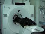
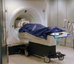
Postmortem volume scanning of a packed human corpse using Multislice Computed Tomography. |
|
|
|
|
 |
Download High-res Image |
|
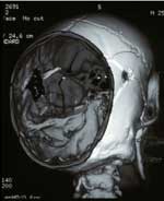
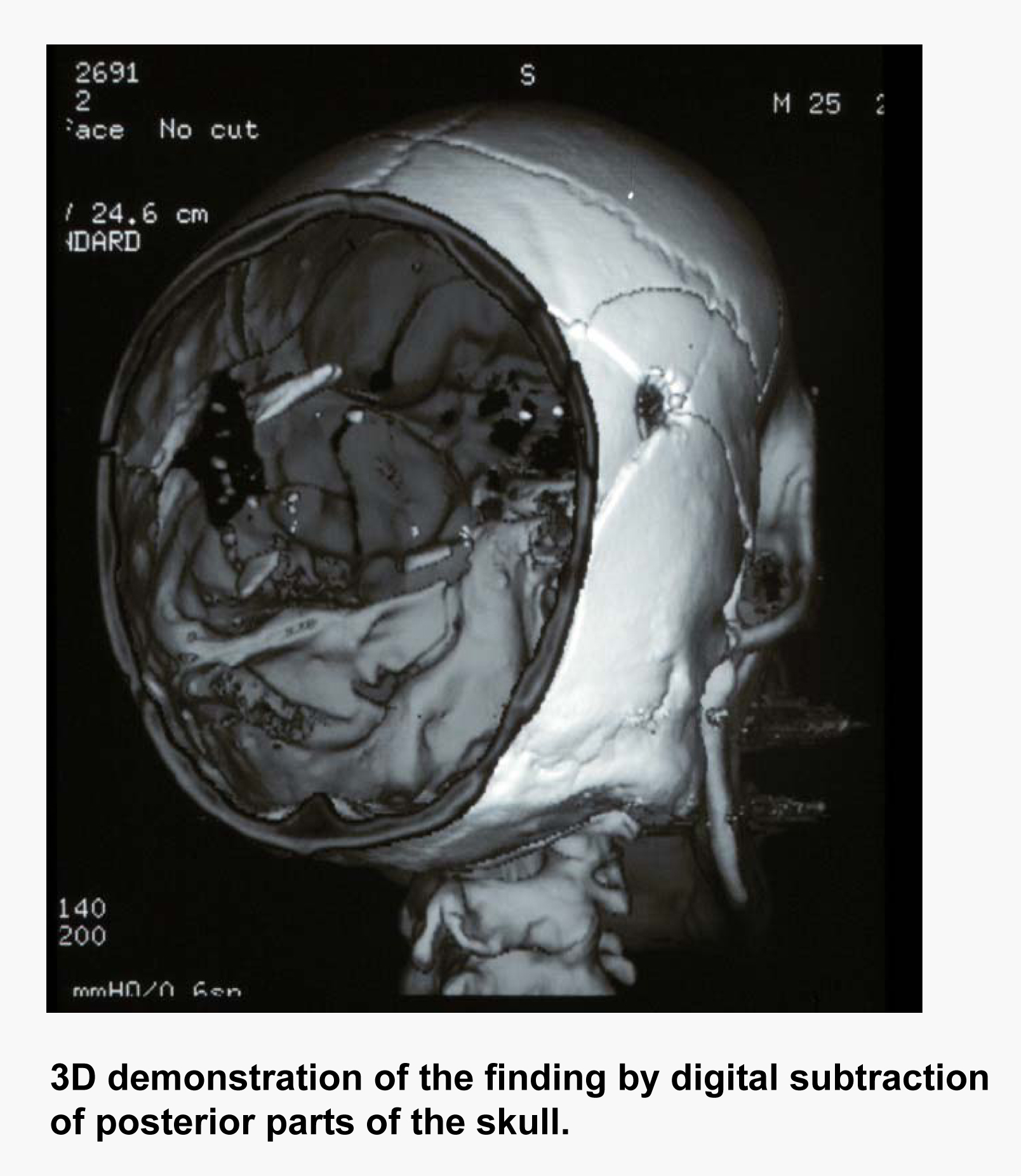
3D demonstration of the finding by digital subtraction of posterior parts of the skull. |
|
|
|
||
 |
|
Download High-res Image |
|
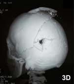
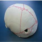
3D reconstruction of the bony structures showing all forensic criteria of an exit wound and additionally the fracture system of the skull. |
Traditional bony preparation / maceration in this case. |
|
|
|
||
 |
 |
Download High-res Image |
|
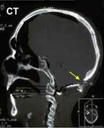
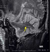
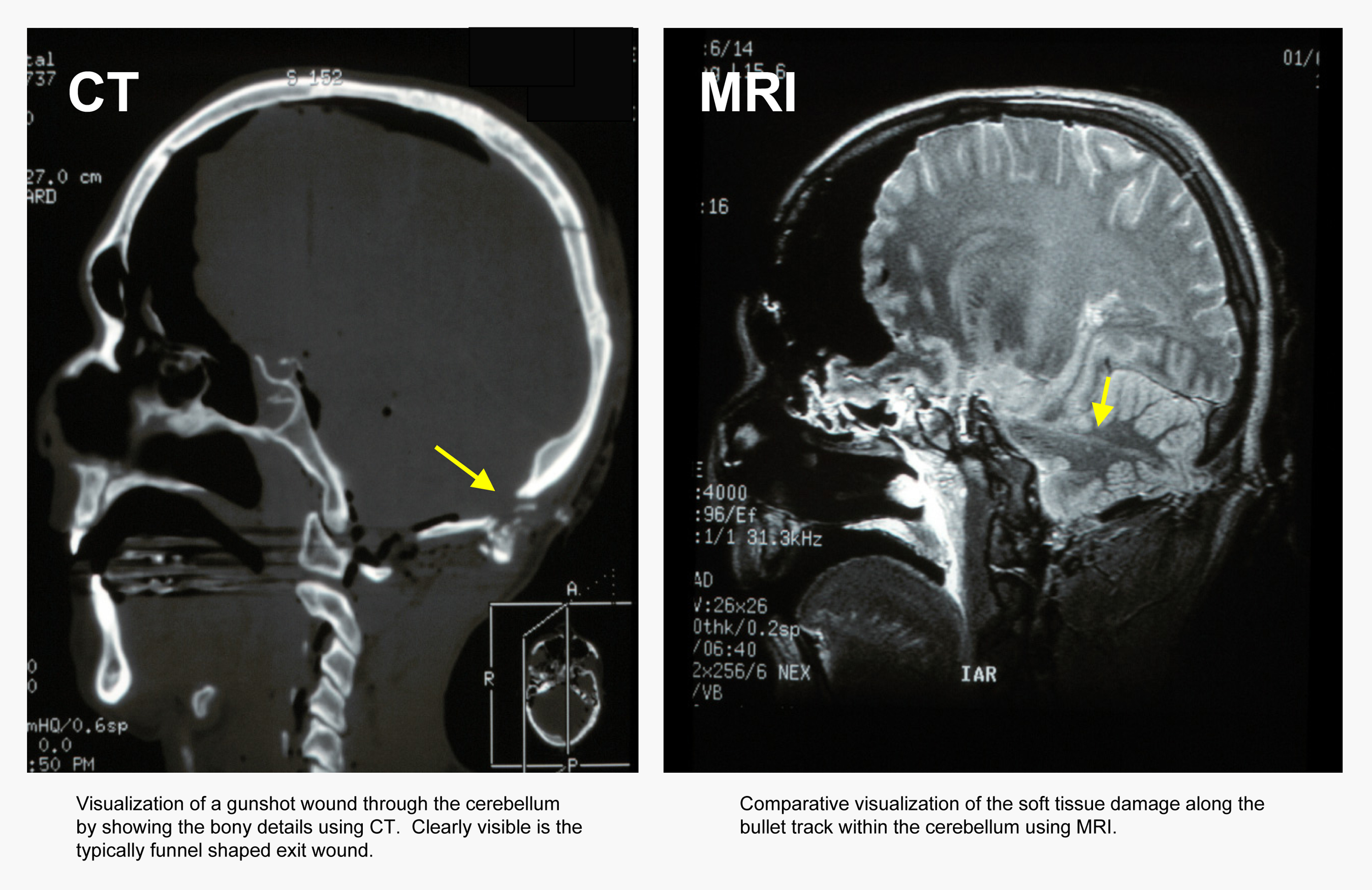
Visualization of a gunshot wound through the cerebellum by showing the bony details using CT. Clearly visible is the typically funnel shaped exit wound. |
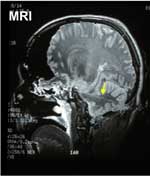
Comparative visualization of the soft tissue damage along the bullet track within the cerebellum using MRI. |
|
|
|
||
 |
 |
|
|
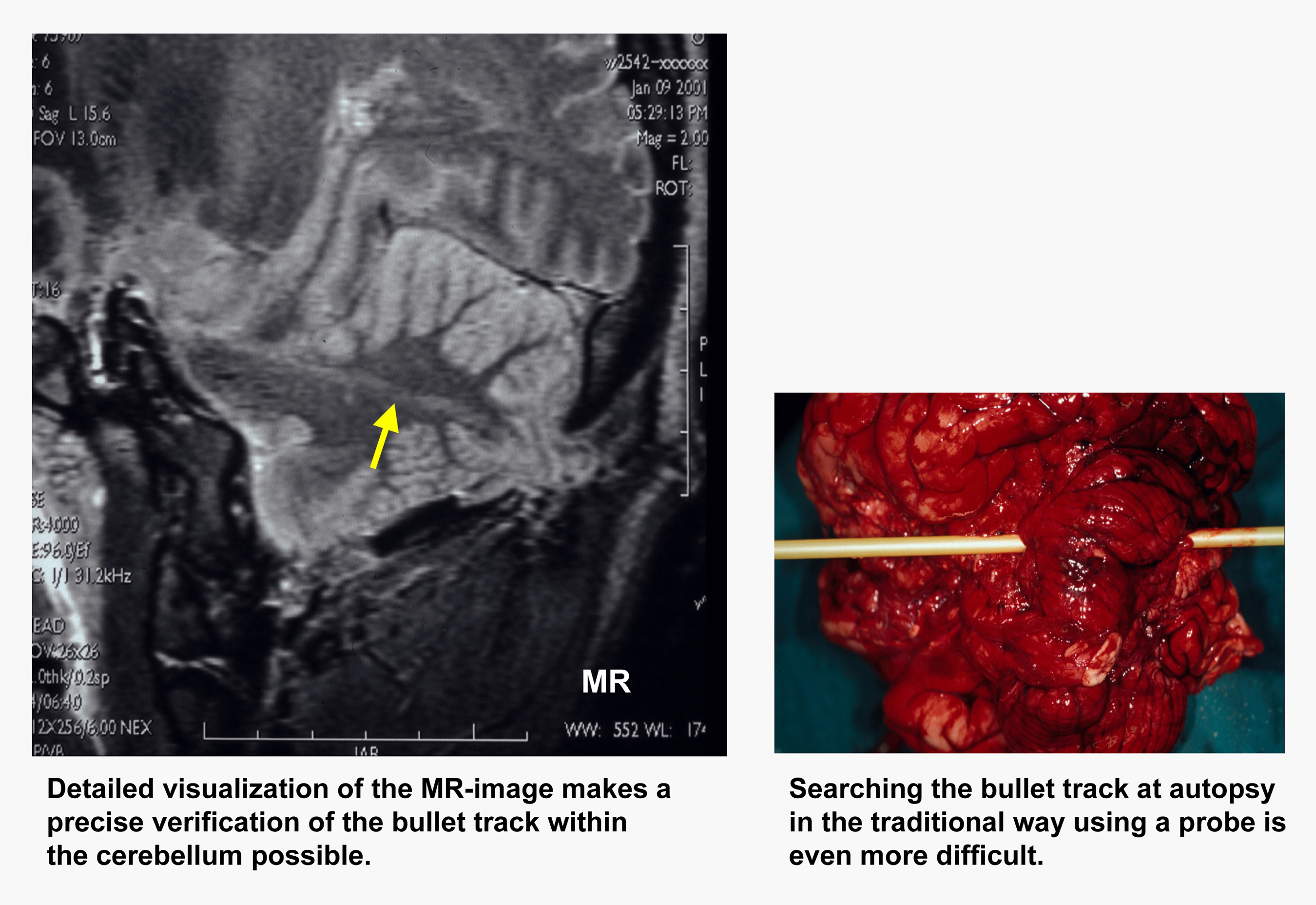
Detailed visualization of the MR-image makes a precise verification of the bullet track within the cerebellum possible. |
Searching the bullet track at autopsy in the traditional way using a probe is even more difficult. |
|
|
|
||
 |
 |
|
|
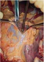
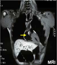
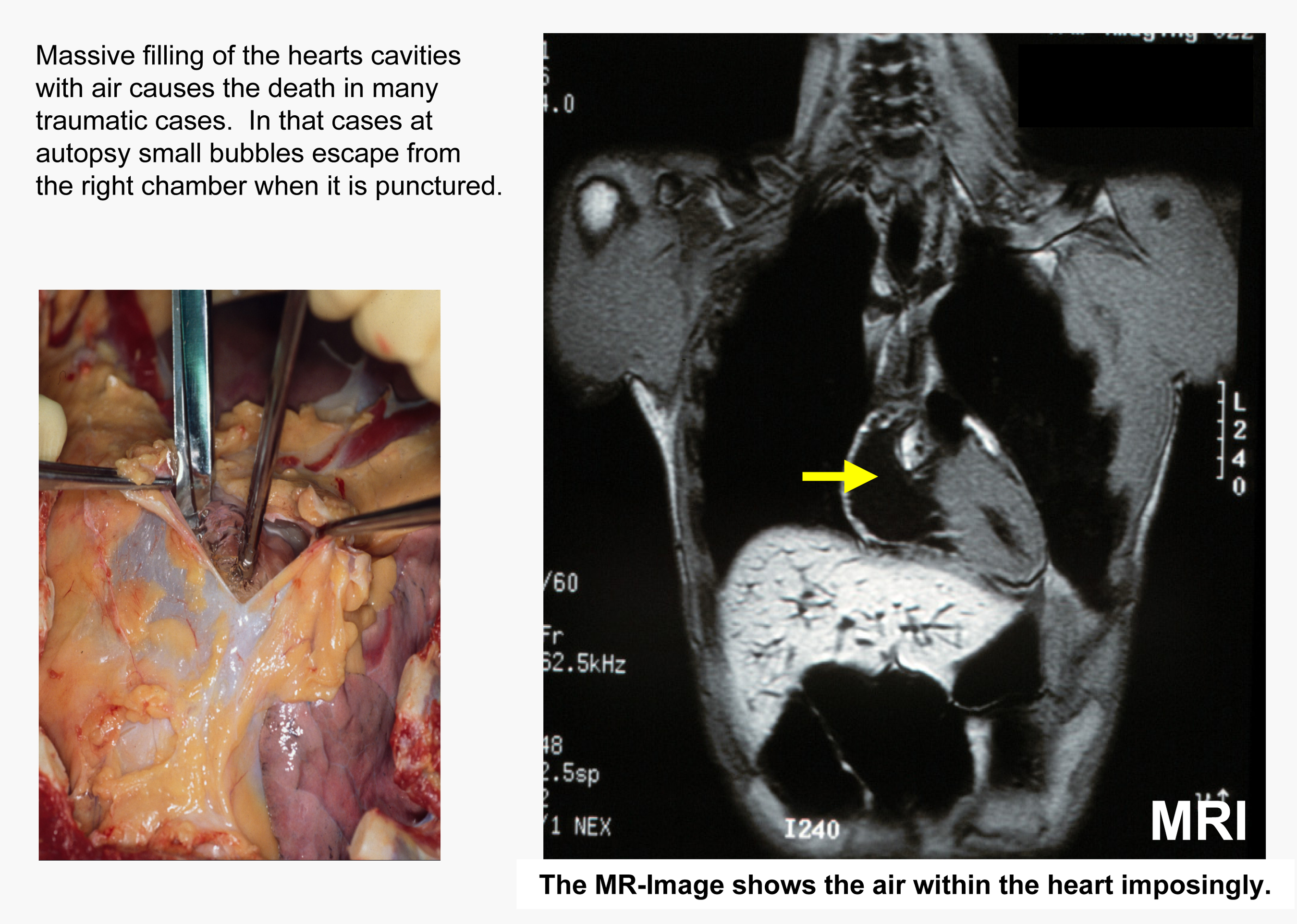
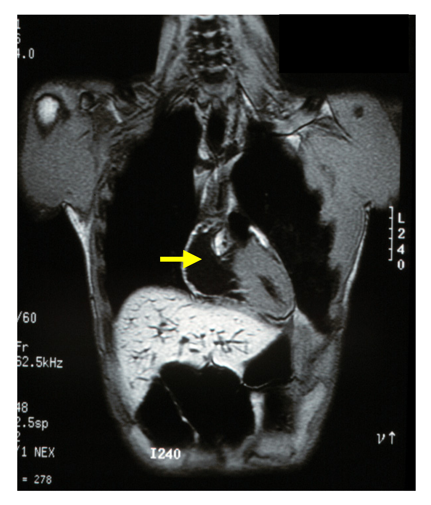
Massive filling of the hearts cavities with air causes the death in many traumatic cases. In that cases at autopsy small bubbles escape from the right chamber when it is punctured. |
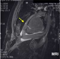
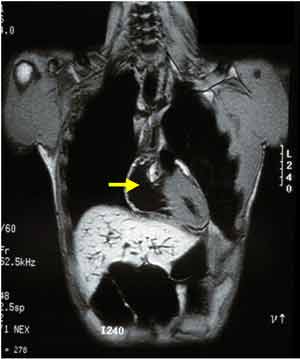
The MR-Image shows the air within the heart imposingly. |
|
|
|
||
 |
 |
|
|
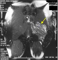
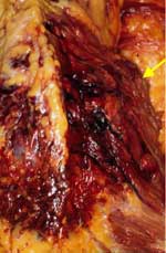
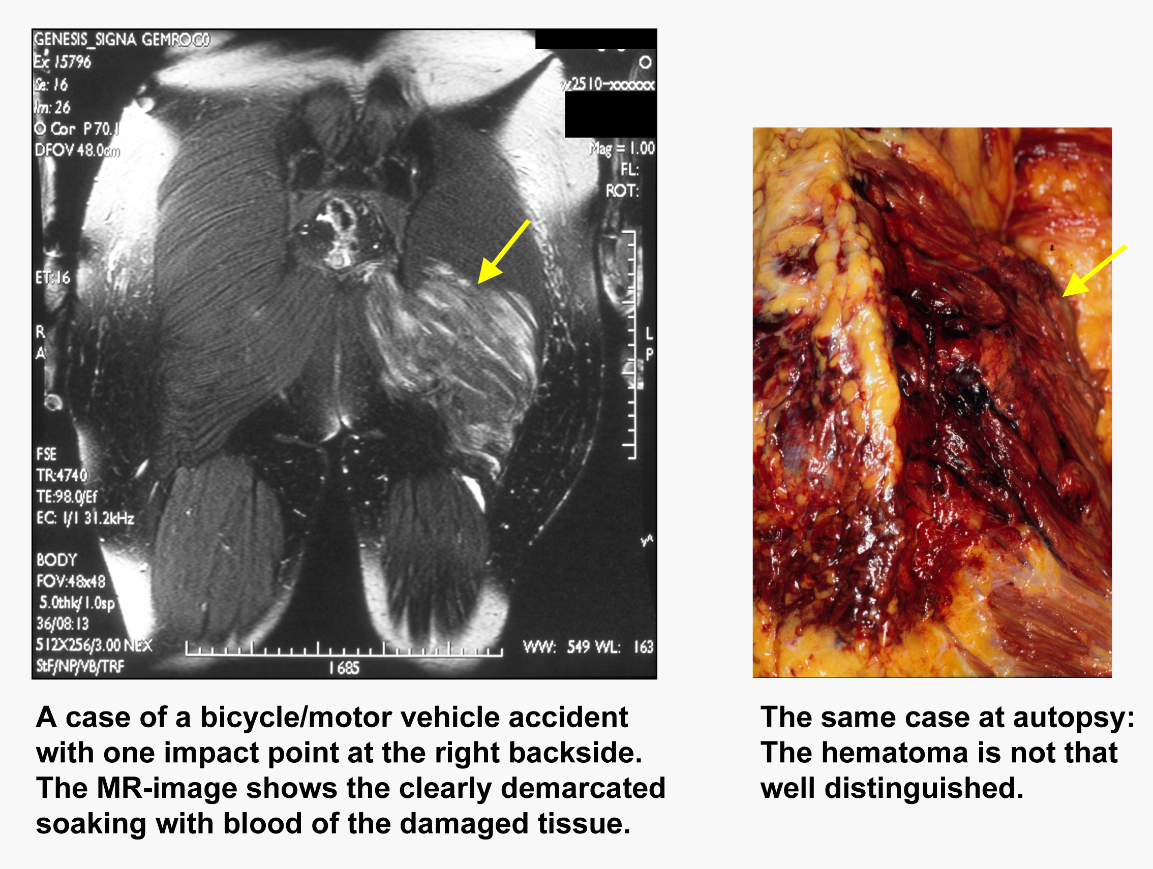
A case of a bicycle/motor vehicle accident with one impact point at the right backside. The MR-image shows the clearly demarcated soaking with blood of the damaged tissue. |
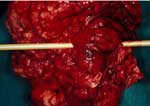
The same case at autopsy: The hematoma is not that well distinguished. |
|
|
|
||
 |
|
|
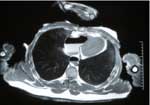
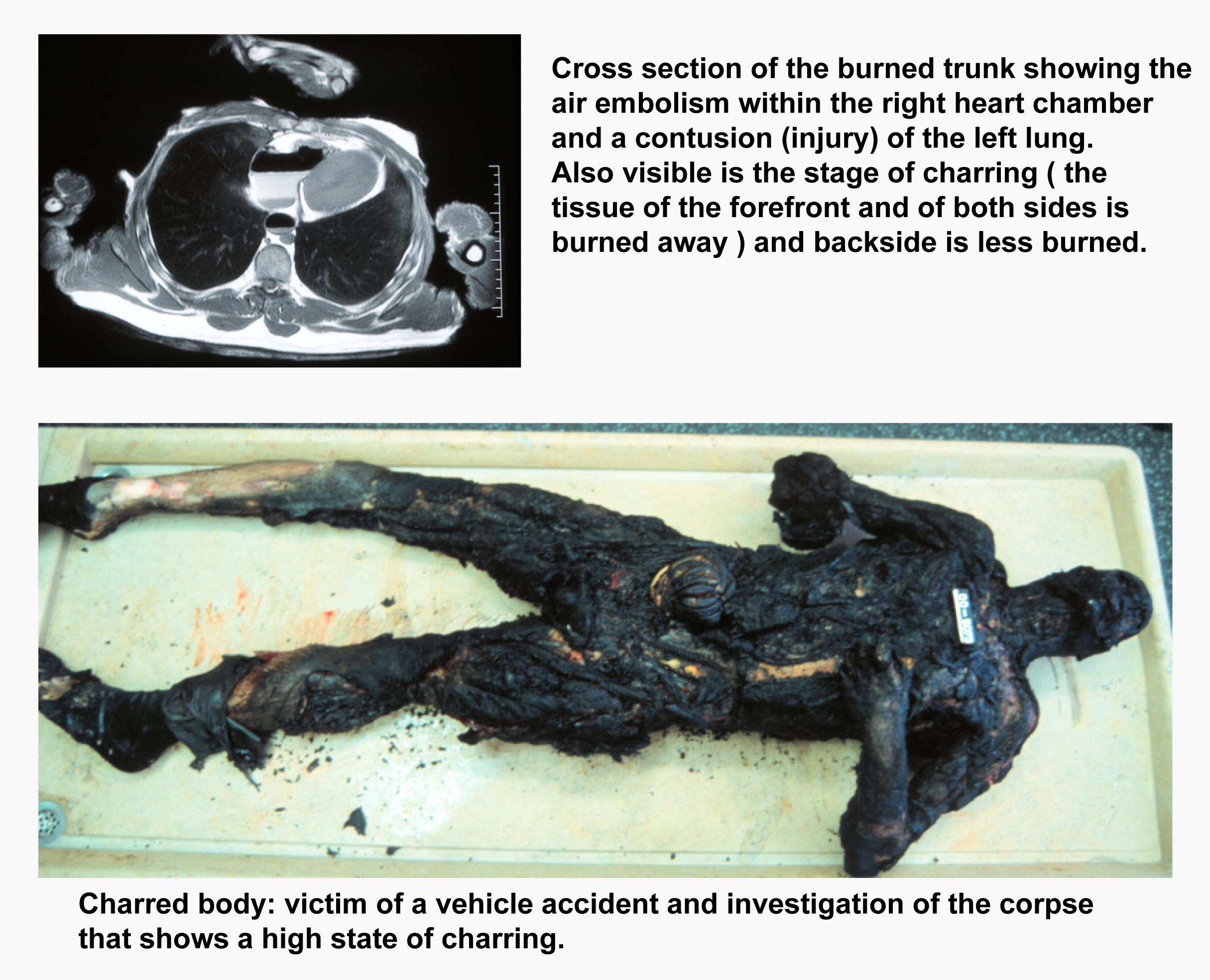
Cross section of the burned trunk showing the air embolism within the right heart chamber and a contusion (injury) of the left lung. Also visible is the stage of charring ( the tissue of the forefront and of both sides is burned away ) and backside is less burned. |
|
 |
|
|
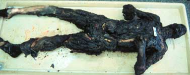
Charred body: victim of a vehicle accident and investigation of the corpse that shows a high state of charring. |
|
|
||
 |
 |
|
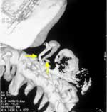
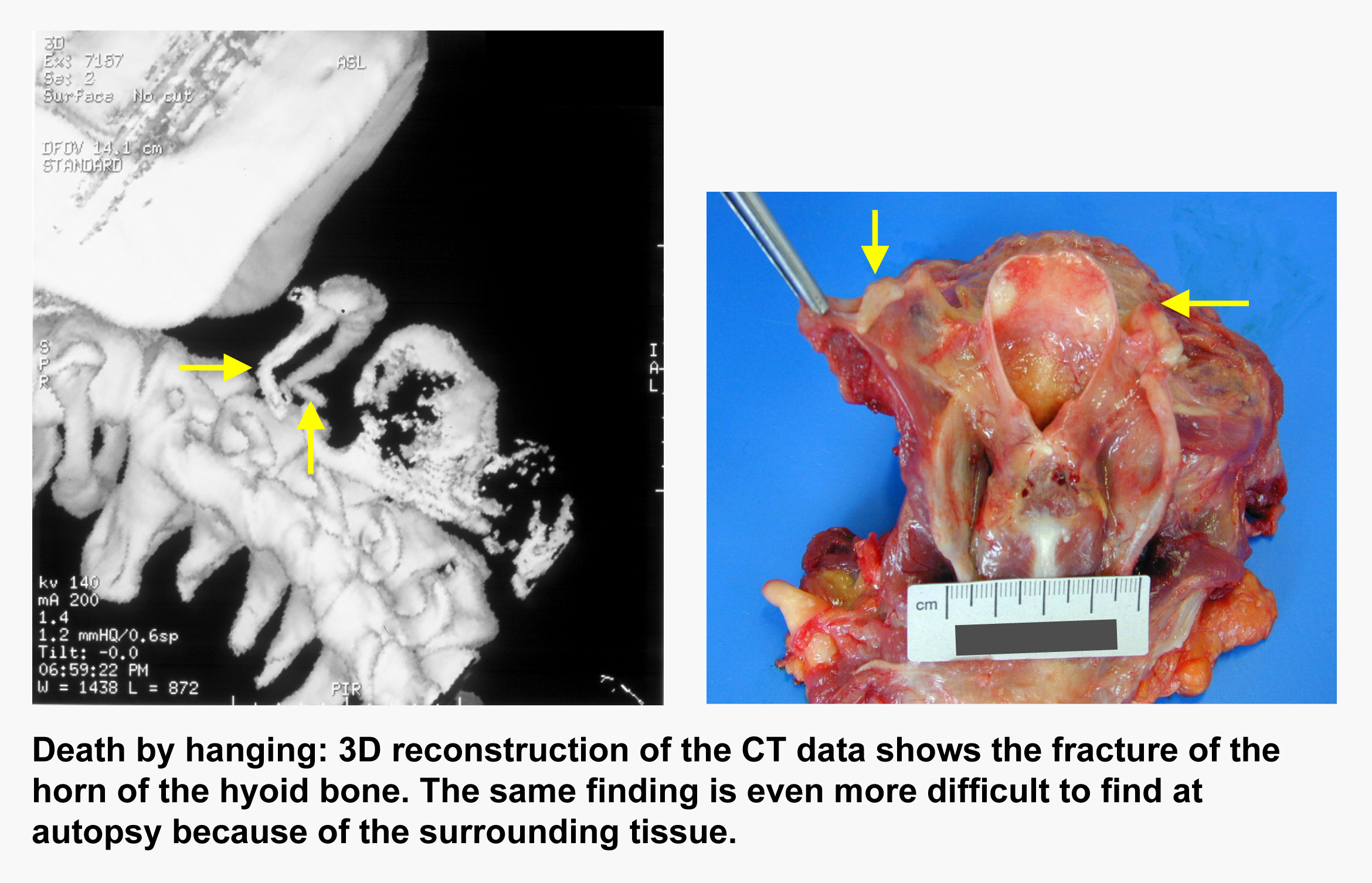
| Death by hanging: 3D reconstruction of the CT data shows the fracture of the horn of the hyoid bone. The same finding is even more difficult to find at autopsy because of the surrounding tissue. | ||
|
|
||
 |
|
|
Body in a body bag during scanning |
|
|
||
 |
 |
|
|
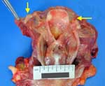
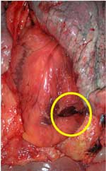
Knife wound to the heart: autopsy and MR finding |
||
|
|
||
 |
|
|
Traumatic, letal air embolism after MVA |
View related abstract:
# # #
 PDF
PDF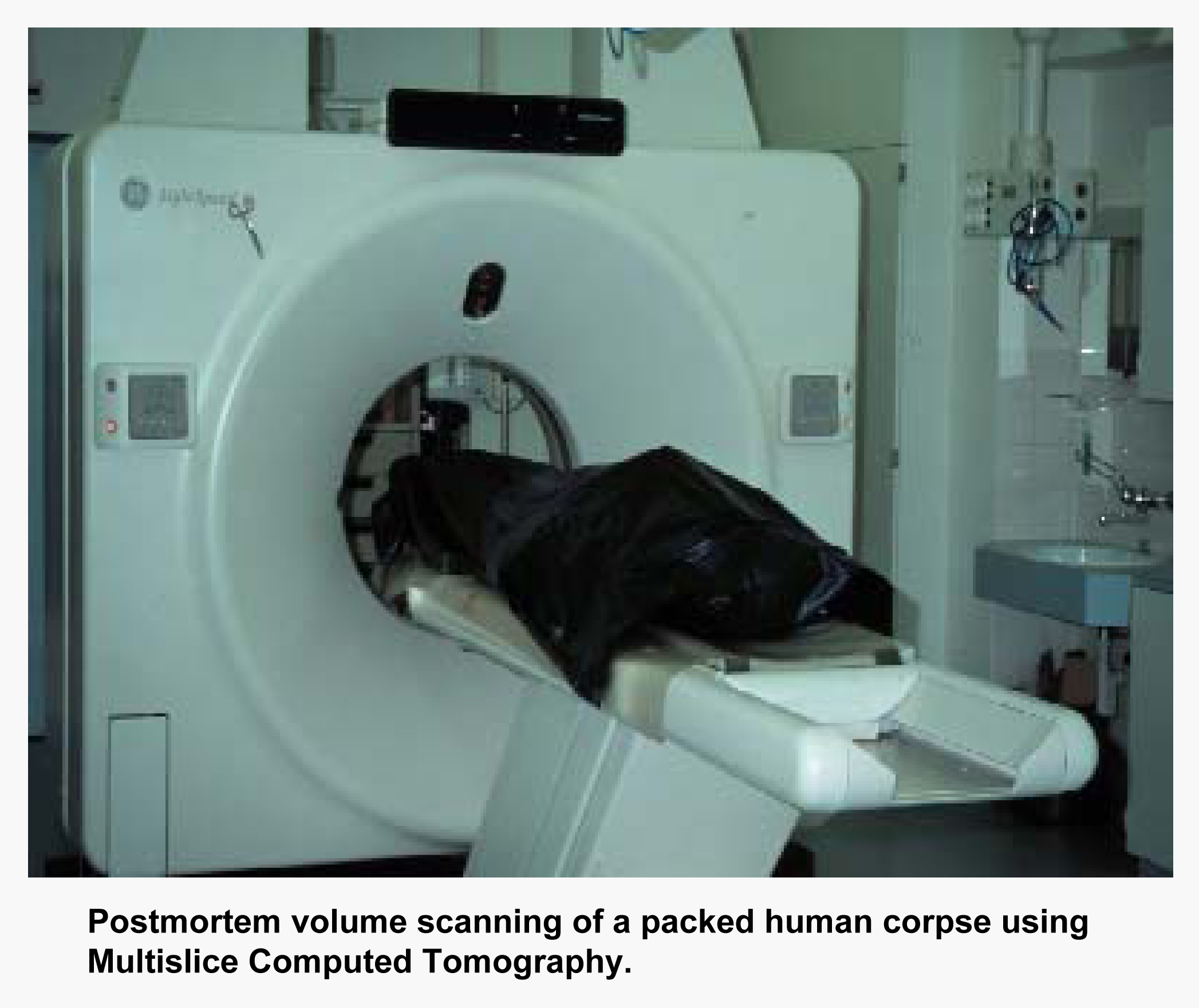