Women Show Persistent Memory Impairment after Concussion
Released: April 28, 2015
At A Glance
- Concussion may affect women more severely than men.
- Functional magnetic resonance imaging exams revealed persistent working memory impairment in women 10 weeks after concussion.
- The findings may indicate a more aggressive treatment protocol is needed for women after concussion.
- RSNA Media Relations
1-630-590-7762
media@rsna.org - Linda Brooks
1-630-590-7738
lbrooks@rsna.org - Emma Day
1-630-590-7791
eday@rsna.org
OAK BROOK, Ill. — Women may have a more difficult time than men in recovering from concussion, according to a new study published online in the journal Radiology.
Concussion, also known as mild traumatic brain injury (MTBI), is a common medical problem affecting cognitive function and quality of life in some individuals.
In most cases, patients who experience MTBI will recover fully, typically within three months. Ten to 15 percent of MTBI patients will continue to experience persistent disabling problems beyond three months, which can include post-traumatic headache, sleep disturbance, loss of balance, memory and other cognitive impairments, fatigue, and mood or affective disorders.

The lasting effects of concussion have received widespread attention in recent years and caused growing concern, particularly among amateur and professional athletes and sports organizations. Several reports have indicated that female athletes suffer concussions at a higher rate than male athletes playing similar sports.
"In clinical practice, more women than men seek medical attention due to persistent symptoms after MTBI at a ratio of almost 2:1," said the study's lead author, Chi-Jen Chen, M.D., from the Department of Diagnostic Radiology and Brain and Consciousness Research Center at Taipei Medical University Shuang-Ho Hospital and superintendent at Chia-Yi Hospital in Taiwan. "We started to wonder whether there might be differences in MTBI outcomes between men and women."
Dr. Chen and colleagues set out to objectively evaluate gender differences in MTBI by using functional magnetic resonance imaging (fMRI) to analyze brain activation patterns during working memory tasks.
"Difficulty with working memory is a commonly reported impairment after MTBI," Dr. Chen said. "Since working memory is important for a wide variety of cognitive skills, compromised working memory could have significant effects on everyday life."
The researchers studied 30 patients with MTBI and 30 control patients. Both groups contained an equal number of men and women. Patients underwent an fMRI exam within one month after injury and a follow-up fMRI exam at six weeks after first scan. All participants underwent neuropsychological tests, including digit span and continuous performance test (CPT). Digit span is a short-term memory test measuring how many numbers a participant can remember in sequence. CPT measures a person's sustained and selective attention and impulsivity.
Initial fMRI results of the MTBI patients showed increased activation in working memory brain circuits in the men and decreased activation in the women, compared to the controls. At follow-up, men with MTBI returned to a normal activation pattern similar to the controls, whereas the women showed persistent hypoactivation, suggesting ongoing working memory problems.
Neuropsychological results showed that among the women, the total digit span score was lower in the MTBI group, compared to the control group.
"These findings provide evidence that female gender may be a risk factor for working memory impairment after MTBI," Dr. Chen said. "If so, more aggressive management should be initiated once MTBI is diagnosed in female patients."
Because fMRI has the potential to detect working memory impairment, predict outcome and monitor treatment effect, incorporation of fMRI into treatment protocol for MTBI may be advisable in the future, according to Dr. Chen. However, he cautions that further study needs to be done to validate these preliminary findings.
"Sex Differences in Working Memory after Mild Traumatic Brain Injury: A Functional MR Imaging study." Collaborating with Dr. Chen were Hui-Ling Hsu, M.D., David Yen-Ting Chen, M.D., Ying-Chi Tseng, M.D., Ying-Sheng Kuo, M.D., Yen-Lin Huang, M.D., Wen-Ta Chiu, M.D., Ph.D., Feng-Xian Yan, M.S., and Wei-Shuan Wang, B.S.
Radiology is edited by Herbert Y. Kressel, M.D., Harvard Medical School, Boston, Mass., and owned and published by the Radiological Society of North America, Inc. (http://radiology.rsna.org/)
RSNA is an association of more than 54,000 radiologists, radiation oncologists, medical physicists and related scientists, promoting excellence in patient care and health care delivery through education, research and technologic innovation. The Society is based in Oak Brook, Ill. (RSNA.org)
For patient-friendly information on fMRI of the brain, visit RadiologyInfo.org.
Images (.JPG and .TIF format)
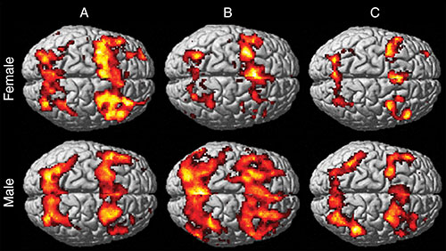
Figure 1. Working memory functional activation in healthy control subjects and patients with MTBI. Surface-rendered projection on a representative atlas brain shows areas of activation in response to increased working memory load. Note the presence of increased activation in bilateral frontal and parietal regions, a finding consistent with activation of working memory circuitry in each group. A: In control subjects, visual comparison of working memory activation patterns shows more activation in women than in men, especially in the frontal region. B: There is substantially less activation seen in female patients with MTBI than in female control subjects, and there is substantially more activation seen in male patients with MTBI than in male control subjects. C: At the 6-week follow-up study, female patients with MTBI showed a persistent hypoactivation pattern, whereas male patients with MTBI showed regression of hyperactivation and demonstrated a return to similar activation levels as those in male control subjects.
High-res (TIF) version
(Right-click and Save As)
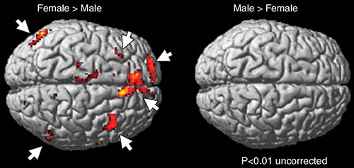
Figure 2. Sex differences of working memory functional activation in healthy control subjects in response to increased working memory load. A voxel-by-voxel comparison of female (left image) and male (right image) control subjects shows more activation (arrows) in female control subjects than in male control subjects.
High-res (TIF) version
(Right-click and Save As)
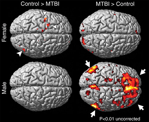
Figure 3. Sex differences of working memory functional activation at initial study of patients with MTBI compared with control subjects in response to increased working memory load. Voxel-by-voxel comparison of initial studies of patients with MTBI and control subjects shows a little more activation in the working memory circuitry (arrowheads) in female control subjects than in female patients with MTBI. However, substantially more activation (arrows) is seen in male patients with MTBI than in male control subjects.
High-res (TIF) version
(Right-click and Save As)