MR Spectroscopy Shows Precancerous Breast Changes in Women with BRCA Gene
Released: March 03, 2015
At A Glance
- A technique that monitors biochemical changes in tissue could improve the management of women at risk of breast cancer.
- Researchers performed 2-D localized correlated spectroscopy (L-COSY) on women carrying the BRCA1 and BRCA2 gene mutations and compared the results with a control group who had no family history of breast cancer.
- L-COSY identified statistically significant biochemical changes in women with BRCA1 and BRCA2 gene mutations compared to controls.
- RSNA Media Relations
1-630-590-7762
media@rsna.org - Linda Brooks
1-630-590-7738
lbrooks@rsna.org - Emma Day
1-630-590-7791
eday@rsna.org
OAK BROOK, Ill. — A magnetic resonance spectroscopy (MRS) technique that monitors biochemical changes in tissue could improve the management of women at risk of breast cancer, according to a new study published online in the journal Radiology.
Many women face a higher risk of breast cancer due to the presence of BRCA gene mutations. A BRCA1 or BRCA2 mutation carrier has approximately a 50 percent risk of developing breast cancer before the age of 50, and the cancer can develop within months of a negative screening by mammography. The risk is so significant that many women with BRCA mutations undergo prophylactic, or preventive mastectomies to avoid getting invasive cancer later in life.
For the new study, researchers assessed 2-D localized correlated spectroscopy (L-COSY) as a noninvasive means to identify biochemical changes associated with a very early stage of cancer development known as the pre-invasive state.
The researchers performed L-COSY on nine women carrying the BRCA1 and 14 women with BRCA2 gene mutations and compared the results with those from 10 healthy controls who had no family history of breast cancer. All the patients underwent contrast enhanced 3-T MRI and ultrasound.
While no abnormality was recorded by MRI or ultrasound, L-COSY MRS identified statistically significant biochemical changes in women with BRCA1 and BRCA2 gene mutations compared to controls. The researchers found multiple distinct cellular changes measurable through L-COSY indicative of premalignant changes in women carrying BRCA gene mutations.
"These changes appear to represent a series of early warning signs that may allow women to make informed decisions as to when and if they have prophylactic mastectomy," said Carolyn Mountford, M.Sc., D.Phil., from the University of Newcastle in Callaghan, Australia, Brigham and Women's Hospital in Boston and the Translational Research Institute in Brisbane.
Study co-author David Clark, M.B.B.S., B.Sc., F.R.A.C.S., from the Breast and Endocrine Centre in Gateshead, New South Wales, Australia, believes the protocol may help guide treatment decisions in women with BRCA mutations. Dr. Clark noted that approximately half the women who have BRCA mutations may not develop breast cancer at all and certainly not before they turn 50 years old, so the spectroscopy technique could be extremely useful.
"We think there are three stages of pre-cancer progression in the breast tissue," he said. "Women at Stage 1 could monitor their breasts with follow-up spectroscopy every six months."
The research team also found evidence that lipid pathways are affected differently in the two different gene mutations, which may help explain why BRCA2 mutation carriers survive longer than BRCA1 carriers.
The study represents the culmination of more than 25 years of work, according to Dr. Mountford. Research on biopsy samples in the 1980s proved the existence of pre-invasive states, but technological improvements were needed before the technique could be applied to clinical MRI scanners.
"It took a multidisciplinary team, including an MR physicist, chemists, radiographers and radiologists to be sure that what we were seeing was not apparent from conventional contrast-enhanced imaging," Dr. Mountford said.
The researchers hope to confirm the findings in larger populations and continue to monitor the women in the study group with the 2-D L-COSY protocol to learn more about the biochemical changes and what they represent.
"Lipid and Metabolite Deregulation in the Breast Tissue of Women Carrying BRCA1 and BRCA2 Genetic Mutations." Collaborating with Drs. Mountford and Clark were Saadallah Ramadan, Ph.D., Jameen Arm, B.Sc., Judith Silcock, R.N., Gorane Santamaria, M.D., Ph.D., Jessica Buck, B.Sc., Michele Roy, M.D., F.R.C.R., Kin Men Leong, M.B.B.S., F.R.A.C.R., Peter Lau, M.B.B.S., F.R.A.C.R., Peter Malycha, M.B.B.S., F.R.A.C.S., F.R.C.S.
Radiology is edited by Herbert Y. Kressel, M.D., Harvard Medical School, Boston, Mass., and owned and published by the Radiological Society of North America, Inc. (http://radiology.rsna.org/)
RSNA is an association of more than 54,000 radiologists, radiation oncologists, medical physicists and related scientists, promoting excellence in patient care and health care delivery through education, research and technologic innovation. The Society is based in Oak Brook, Ill. (RSNA.org)
For patient-friendly information on breast imaging, visit RadiologyInfo.org.
Images (.JPG and .TIF format)

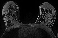
Figure 1. Unenhanced image shows region of interest placement in a 30-year-old control subject. This is the spectroscopic voxel location from which 2D localized COSY data were collected.
High-res (TIF) version
(Right-click and Save As)

Figure 2. Diagram of a fatty acyl chain, with the associated spectroscopic cross peaks (A–G’) labeled.
High-res (TIF) version
(Right-click and Save As)

Figure 3. Diagram of the structure of a cholesterol molecule, with the methyl group used for spectroscopic detection in bold.
High-res (TIF) version
(Right-click and Save As)

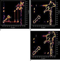
Figure 4. Typical localized COSY spectra in three women with the cross peaks assigned (A–G’) as per Figure 2. Images in (a) healthy 55-year-old con¬trol subject, (b) apparently healthy 56-year-old woman with BRCA1 mutation, and (c) apparently healthy 58-year-old woman with BRCA2 mutation.
High-res (TIF) version
(Right-click and Save As)