RSNA Press Release
- MR-guided focused ultrasound (MRgFUS) ablation is a noninvasive breast cancer treatment technique.
- Post-surgical evaluation confirmed the absence of residual disease in the treatment area in 83 percent of patients.
- None of the patients in the study experienced significant complications.
MR-guided Ultrasound Offers Noninvasive Treatment for Breast Cancer
Released: December 4, 2013
| Media Contacts: | RSNA Newsroom | 1-312-949-3233 |
| Before 11/30/13 or after 12/5/13: | RSNA Media Relations: | 1-630-590-7762 |
| |
Linda Brooks 1-630-590-7738 lbrooks@rsna.org |
Maureen Morley 1-630-590-7754 mmorley@rsna.org |
CHICAGO – A technique that uses focused ultrasound under magnetic resonance (MR) guidance to heat and destroy tumors may offer a safe and effective treatment for breast cancer, according to research being presented today at the annual meeting of the Radiological Society of North America (RSNA).
MR-guided focused ultrasound (MRgFUS) ablation is a noninvasive technique that requires no incision or puncture to perform. Instead, it uses the acoustic energy from high-intensity focused ultrasound to remove, or ablate, diseased tissue. Continuous MRI is used to locate the lesions and monitor the temperature change during the ablation process.

Primary advantages of MRgFUS over other breast cancer treatments are that it is a noninvasive, outpatient procedure offering a quick recovery time, and that it provides precise measurement of temperature changes during the procedure.
"In the treatment stage, we are able to precisely visualize where the energy is having an effect and to measure exactly the rise in temperature," said Alessandro Napoli, M.D., Ph.D., assistant professor of radiology at Sapienza University in Rome. "Temperature monitoring is particularly important, since too low a temperature is ineffective and too high a temperature may be dangerous."
Dr. Napoli and colleagues assessed the safety and efficacy of MRgFUS in 12 patients with invasive ductal breast cancer before surgical removal of the cancer and biopsy of the lymph nodes. They used 3T MRI to confirm the presence and treatable location of cancerous lesions. The patients then underwent single-session MRgFUS treatment. Researchers evaluated treatment efficacy through post-surgery pathology.
None of the patients experienced significant complications during or immediately after the procedure. In 10 of the 12 patients, MRI showed no enhancement in the treatment area after the procedure. Post-surgery histological evaluation confirmed the absence of residual disease in the treatment area in those 10 patients.
"This procedure allows for safe ablation of breast cancer," Dr. Napoli said. "At pathology, no significant viable tumor was found in the specimens from these 10 patients."
In the other two cases, treatment failed due to transducer malfunction, and the pathologist observed residual tumor in the samples.
According to Dr. Napoli, MRI guidance is crucial for correct identification of lesions, treatment planning and real-time control during the procedure. Specifically, monitoring with MRI allows for efficient deposit of energy into the region of treatment at the correct range of between 60 degrees and 70 degrees Celsius (approximately 140 to 158 degrees Fahrenheit).
"This is carried out by a special sequence that is called MR thermometry," Dr. Napoli said. "Only MRI presently has the ability to determine, in real time, fine temperature quantification."
While the initial results are promising, Dr. Napoli said more research will be needed before the approach can be adopted as a stand-alone treatment for breast cancer.
Co-authors are Luisa Di Mare, M.D., Federica Pediconi, M.D., Michele Anzidei, M.D., Vincenzo Noce, M.D., and Carlo Catalano, M.D.
# # #
Note: Copies of other RSNA 2013 news releases and electronic images will be available online at RSNA.org/press13 beginning Monday, Dec. 2.
RSNA is an association of more than 53,000 radiologists, radiation oncologists, medical physicists and related scientists, promoting excellence in patient care and health care delivery through education, research and technologic innovation. The Society is based in Oak Brook, Ill. (RSNA.org)
Editor's note: The data in these releases may differ from those in the published abstract and those actually presented at the meeting, as researchers continue to update their data right up until the meeting. To ensure you are using the most up-to-date information, please call the RSNA Newsroom at 1-312-949-3233.
For patient-friendly information on breast cancer diagnosis and treatment, visit RadiologyInfo.org.
| Abstract: |
Video clips (.mp4 format)
 Video 1. Footage showing an animation of MR-guided focused ultrasound (MRgFUS) ablation. |
Images (.JPG format)
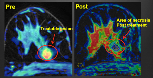 Figure 1. PRE: Pre-treatment transverse MR image obtained with perfusion technique shows an enhancing lesion of 1.2 cm (circled) in the upper quadrants of the right breast; the lesion shows with irregular margin and appears color-coded in red due to the wash-out pattern (malignancy). POST: Same MR image technique obtained post treatment (10 days): Absence of enhancement was seen at perfusion color-coded image after non-invasive thermal ablation with MR guided high intensity focused ultrasound; the black hole stands for necrosis nicely demonstrating also the absence of residual tumor. High-res (TIF) version (Right-click and Save As) |
 PDF
PDF