RSNA Press Release
- Radiation dose to areas of the body near the breast during mammography is very low.
- Researchers measured the dose received by the thyroid gland, salivary gland, sternum, uterus and the lens of the eye during screening digital mammography.
- Thyroid shields are unnecessary during mammography and could actually result in an increased radiation dose to patients.
Scatter Radiation from Mammography Presents No Cancer Risk
Released: November 27, 2012
| Media Contacts: | RSNA Newsroom | 1-312-949-3233 |
| Before 11/24/12 or after 11/29/12: | RSNA Media Relations: | 1-630-590-7762 |
| |
Linda Brooks 1-630-590-7738 lbrooks@rsna.org |
Maureen Morley 1-630-590-7754 mmorley@rsna.org |
CHICAGO—The radiation dose to areas of the body near the breast during mammography is negligible, or very low, and does not result in an increased risk of cancer, according to a study presented today at the annual meeting of the Radiological Society of North America (RSNA). The results suggest that the use of thyroid shields during mammography is unnecessary.

"Thyroid shields can impede good mammographic quality and, therefore, are not recommended during mammography," said Alison L. Chetlen, D.O., assistant professor of radiology at Penn State Hershey Medical Center.
During mammography, some X-rays scatter away from the primary beam in the breast and spread outward in different directions. Although this scatter radiation is much weaker than the primary beam, there has been concern that women exposed to it during mammography could face an increased risk of cancer, especially in radiosensitive areas like the thyroid gland.
To better understand the potential impact of scatter radiation, Dr. Chetlen and colleagues set out to measure the dose received by the thyroid gland, salivary gland, sternum, uterus and the lens of the eye during screening digital mammography. Each of the 207 women in the study group wore six optically stimulated luminescent dosimeters—a device used to measure an absorbed dose of ionizing radiation—while undergoing two-view screening mammography.
Analysis of the dosimeters by a medical physicist immediately after the exam revealed that the doses to the various areas outside of the breast ranged from negligible to very low.
Absorbed radiation dose is measured in a unit called a milligray (mGy). The average estimated organ dose to the salivary gland was 0.05 mGy. The average estimated organ dose to the thyroid gland was 0.05 mGy. These doses are only a fraction of the radiation people are exposed to from natural background sources, such as cosmic radiation and radionuclides in the ground. In fact, all areas except for the sternum received less than 2 percent of annual background radiation dose.
Measured dose to the bridge of the eye and umbilicus was negligible, indicating no increased risk to the patient of cataracts or interference with normal embryonic development in early pregnancy.
"The risk of cancer induction at these low levels is indistinguishable from background incidence of cancer due to other sources," Dr. Chetlen said.
The findings are particularly important in light of a recent increase in the incidence of thyroid cancer, one of the most radiosensitive of all cancers. The number of thyroid cancer diagnoses in women nearly doubled from 2000 to 2008, leading some to suspect that mammography may be a contributing factor and that women should wear lead thyroid shields during exams, an idea that Dr. Chetlen and other mammography experts strongly discourage.
Based on the extremely low scatter radiation dose to the thyroid—equivalent to just a few minutes of background radiation—thyroid shields are unnecessary during mammography. In addition, the researchers warn that use of thyroid shields could result in an increased radiation dose to patients.
"A thyroid shield gets in the way of the exam and can actually cause an increase in radiation dose by necessitating repeat exams," Dr. Chetlen said.
Dr. Chetlen also pointed out that the thyroid gland is far less radiosensitive after age 30. The American Cancer Society and other organizations recommend that women have mammography screening once every year, beginning at age 40.
"In the age group eligible for screening, the thyroid gland is not very radiosensitive," Dr. Chetlen said.
Coauthors are Steven King, M.S., Karen Brown, C.H.P., D.A.B.R., Brian Lorah, Susann Schetter, D.O., Claudia Kasales, M.D., Shelley Tuzzato, R.T.R.M., and Shelly Rambler, R.T.R.M.
# # #
Note: Copies of RSNA 2012 news releases and electronic images will be available online at RSNA.org/press12 beginning Monday, Nov. 26.
RSNA is an association of more than 50,000 radiologists, radiation oncologists, medical physicists and related scientists, promoting excellence in patient care and health care delivery through education, research and technologic innovation. The Society is based in Oak Brook, Ill. (RSNA.org)
Editor's note: The data in these releases may differ from those in the published abstract and those actually presented at the meeting, as researchers continue to update their data right up until the meeting. To ensure you are using the most up-to-date information, please call the RSNA Newsroom at 1-312-949-3233.
For patient-friendly information on mammography, visit RadiologyInfo.org.
| Abstract: |
Video clips
- .mp4 format
- Video clip (36,838 Kbyte)
- Video clip (29,438 Kbyte)
- Video clip (32,837 Kbyte)
- Video clip (45,336 Kbyte)
- Video clip (20,350 Kbyte)
- Video clip (21,278 Kbyte)
Images (.JPG format)
 Figure 1. Photograph of optically stimulated luminescence (OSL) dosimeters. High-res (TIF) version (Right-click and Save As) |
 Figure 2. Photograph of a thyroid shield. High-res (TIF) version (Right-click and Save As) |
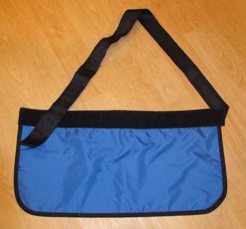 Figure 3. Photograph of a pelvic lead shield. High-res (TIF) version (Right-click and Save As) |
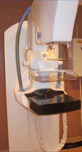 Figure 4. Photograph of a GE Medical Systems Senographe Essential mammography machine used in this study. High-res (TIF) version (Right-click and Save As) |
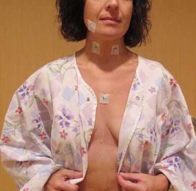 Figure 5. Prior to the study, six optically stimulated luminescence (OSL) dosimeters were taped to the patient’s skin at the following locations: right thyroid lobe, left thyroid lobe, right submandibular gland, midline between eyes on the bridge of nose (not pictured), mid-sternum, and 2 cm caudal to umbilicus (not pictured). Upon completion of four standard screening views the dosimeters were removed and stored in a low background area with a control dosimeter until readout. High-res (TIF) version (Right-click and Save As) |
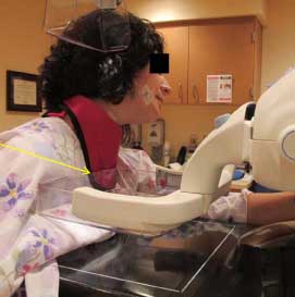 Figure 6. Depending on body habitus of the patient, the thyroid shield could overlie the upper inner breast and findings could be missed because they would not be imaged (arrow). High-res (TIF) version (Right-click and Save As) |
 PDF
PDF