Common Knee Surgery May Lead to Arthritis and Cartilage Loss
Released: December 03, 2014
At A Glance
- Surgery to repair meniscal tears in the knee may accelerate the onset of osteoarthritis and cartilage loss.
- Meniscal surgery is one of the most commonly performed orthopedic procedures.
- In the study, 100% of knees that had undergone surgery went on to develop arthritis, compared to 59% of those that had meniscal damage but did not have surgery.
- RSNA Media Relations
1-630-590-7762
media@rsna.org - Maureen Morley
1-630-590-7754
mmorley@rsna.org - Linda Brooks
1-630-590-7738
lbrooks@rsna.org
CHICAGO – A popular surgery to repair meniscal tears may increase the risk of osteoarthritis and cartilage loss in some patients, according to research presented today at the annual meeting of the Radiological Society of North America (RSNA). The findings show that the decision for surgery requires careful consideration in order to avoid accelerated disease onset, researchers said.

The new study focused on the meniscus, a wedge-shaped piece of cartilage in the knee that acts as a shock absorber between the femur, or thighbone, and tibia, or shinbone. The two menisci in each knee also play an important role in joint stability. Meniscal tears are among the most common knee injuries, and surgery is often performed to alleviate pain.
"Meniscal surgery is one of the most common orthopedic procedures performed to alleviate pain and improve joint function," said Frank W. Roemer, M.D., from Boston University School of Medicine in Boston and the University of Erlangen-Nuremberg in Erlangen, Germany. "However, increasing evidence is emerging that suggests meniscal surgery may be detrimental to the knee joint."
For the study, Dr. Roemer and colleagues examined data from the Osteoarthritis Initiative, a large, ongoing observational study of knee osteoarthritis incidence and progression. Patients in the study were on average 60.2 years old and predominantly overweight, with a mean body mass index (BMI) of 28.3. Approximately two-thirds of the patients were women.
The researchers studied magnetic resonance imaging (MRI) exams of 355 knees that developed osteoarthritis during a five-year period, and a control group that was matched for age, gender, arthritic severity in both knees and BMI. Of all knees, 31 underwent meniscal surgery during the year prior to the arthritis diagnosis, and 280 knees had signs of meniscal damage on MRI but did not have surgery. Also part of the analysis were control cases with no meniscal damage. The researchers assessed the risk of developing arthritis and cartilage loss during the following year for the different groups.
"We found that patients without knee osteoarthritis who underwent meniscal surgery had a highly increased risk for developing osteoarthritis and cartilage loss in the following year compared to those that did not have surgery, regardless of presence or absence of a meniscal tear in the year before," Dr. Roemer said.
All 31 of the knees that underwent meniscal surgery during the prior year developed osteoarthritis, compared with 165 (59 percent) of the knees with meniscal damage that didn't have surgery. In addition, cartilage loss was much more common among knees that had undergone surgery: 80.8 percent of knees with surgery showed cartilage loss, compared with 39.5 percent of knees with meniscal damage and no surgery.
An alternative to surgery is conservative management. In conservative management, physical therapy is prescribed to help maintain and restore muscle strength and range of motion. Symptoms are commonly treated with ice and non-steroidal anti-inflammatory medications.
"The indications for meniscal surgery might need to be discussed more carefully in order to avoid accelerated knee joint degeneration," Dr. Roemer said.
Co-authors on the study are Ali Guermazi, M.D., Ph.D., C. Kent Kwoh, M.D., Michael Hannon, M.A., Jason Grago, M.A., and David J. Hunter, M.D., Ph.D.
Note: Copies of RSNA 2014 news releases and electronic images will be available online at RSNA.org/press14 beginning Monday, Dec. 1.
RSNA is an association of more than 54,000 radiologists, radiation oncologists, medical physicists and related scientists, promoting excellence in patient care and health care delivery through education, research and technologic innovation. The Society is based in Oak Brook, Ill. (RSNA.org)
Editor's note: The data in these releases may differ from those in the published abstract and those actually presented at the meeting, as researchers continue to update their data right up until the meeting. To ensure you are using the most up-to-date information, please call the RSNA Newsroom at 1-312-791-6610.
For patient-friendly information on MRI of the knee, visit RadiologyInfo.org.
Images (.JPG and .TIF format)
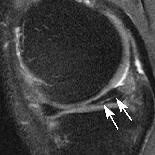
Figure 1A. Examples of meniscal tears and status after surgery. A. Sagittal intermediate-weighted fat-saturated image shows a typical horizontal-oblique meniscal tear of the posterior horn reaching the undersurface of the meniscus (arrows).
High-res (TIF) version
(Right-click and Save As)
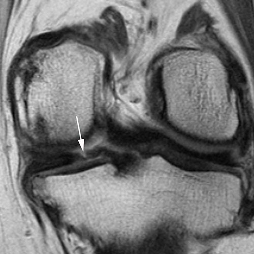
Figure 1B. Examples of meniscal tears and status after surgery. B. Coronal proton density-weighted image shows another example of a tear, which represents a vertical tear of the posterior horn of the medial meniscus (arrow).
High-res (TIF) version
(Right-click and Save As)
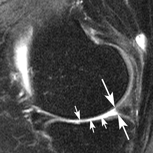
Figure 1C. Examples of meniscal tears and status after surgery. C. Sagittal intermediate-weighted fat-saturated image post surgery shows substance loss of the posterior horn of the medial meniscus (large arrows) and concomitant cartilage loss at the central medial tibia and femur (small arrows).
High-res (TIF) version
(Right-click and Save As)
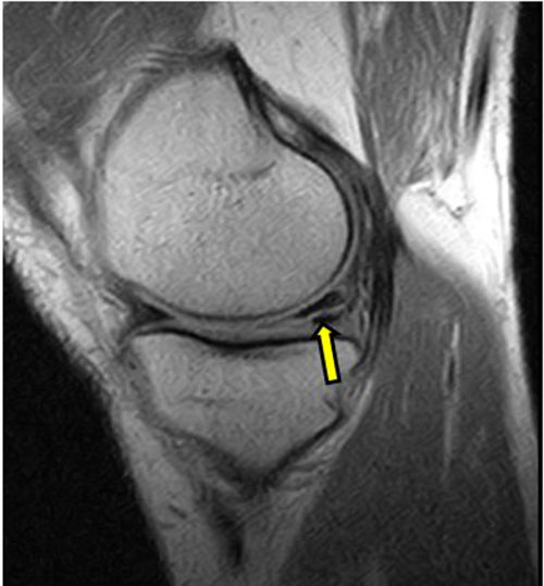
Figure 2. A horizontal tear of the posterior part of the meniscus of the knee.
High-res (TIF) version
(Right-click and Save As)